Your vital organs—screened
Scan your body for potential cancer and 500+ conditions in up to 13 organs.


Our scan is designed to


"A large part of the credit for this great prognosis goes to early detection: given that the tumor was found so early, it was easier to remove surgically, and any spread is unlikely"

Most people diagnosed with cancer twice can’t say cancer and lucky in the same sentence. I'm so thankful to have caught these cancers early.


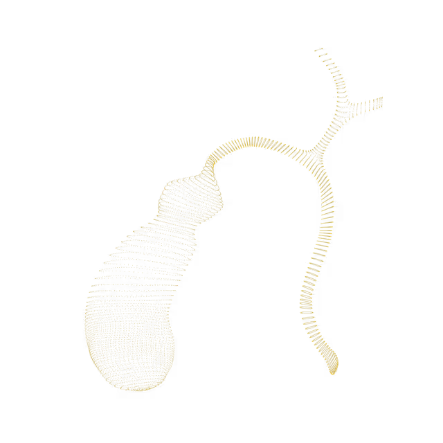
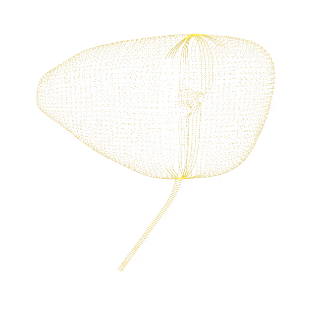
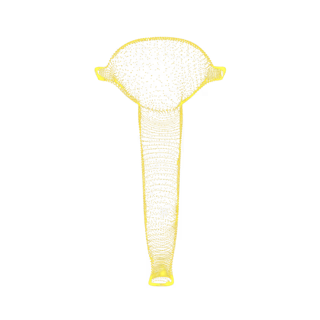
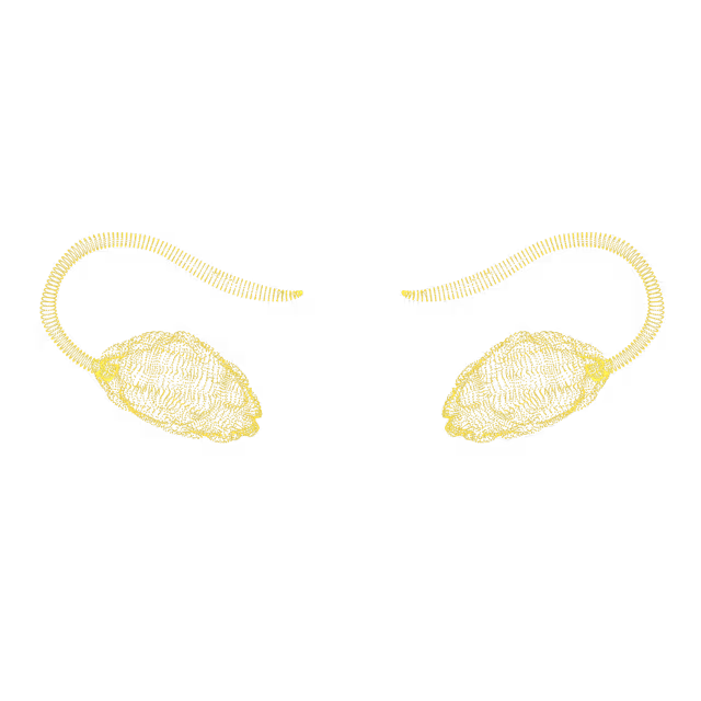
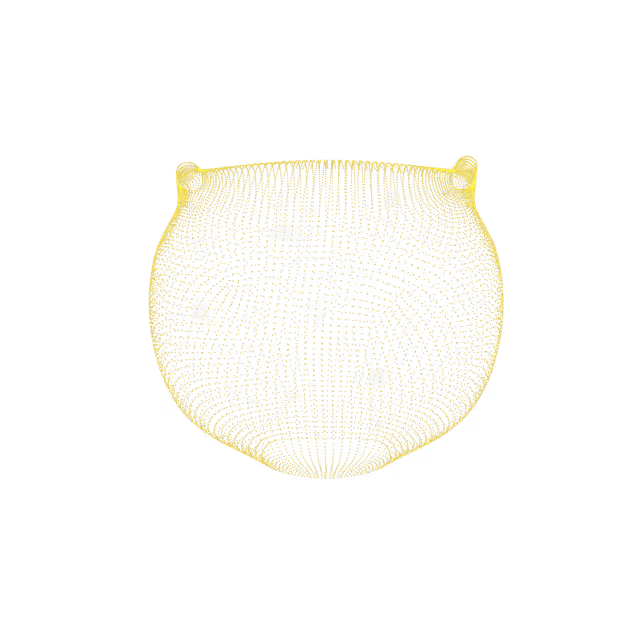

The gallbladder is a small, pear-shaped organ on the right side of the abdomen, just beneath the liver. The gallbladder holds a digestive fluid called bile that is released into the small intestine. Gallstones (cholelithiasis) can form when digestive fluid deposits and hardens. Cholecystitis is an inflammation of the gallbladder.Based on your MRI images, there is no evidence of acute inflammation of the gallbladder but there is evidence of gallstones.
The gallbladder contracts ("shrinks") when it releases bile, and then relaxes, returning to its usual size - this physiologic process can show a "contracted gallbladder" on radiologic imaging. Sometimes, a contracted gallbladder can be caused by chronic inflammation (e.g. symptomatic gallstones) causing gallbladder scarring and a permanently smaller-than-usual gallbladder.
The extrahepatic bile duct is a tube that is outside the liver and carries bile from the liver and gallbladder to the small intestine. Dilatation is the expansion or widening of the duct. Dilatation of the extrahepatic bile duct can be due to several causes including early gallstones in the bile duct, obstruction of the common bile duct at the sphincter of Oddi, pregnancy, the presence of a cyst in the bile duct, and/or drugs (e.g. chronic opioid use). Symptoms, if present, could include right upper quadrant pain and/or jaundice.
Bile duct cysts are fluid-filled pockets that form along the biliary duct (the tube that carries bile from the liver and gallbladder into the intestines). It is not known what causes bile duct cysts to form. These cysts can sometimes cause abdominal pain, jaundice (yellowing of the skin) and/or abdominal mass. Bile duct cysts can be a pre-cancer finding, so discuss this with your primary care provider and a gastroenterologist or hepatologist (liver specialist) for further evaluation and management.
The gallbladder, which is part of the biliary tree, is a small organ located under the liver that stores bile, a substance that helps to break down fats. A cyst is a fluid-filled pocket. It is not known what causes gallbladder cysts to form. These cysts can sometimes cause abdominal pain, jaundice (yellowing of the skin or eyes) and/or an abdominal mass. Biliary tree cysts can be a precancerous finding, so discuss this with your primary care provider and a gastroenterologist or hepatologist (liver specialist) for continued follow-up and surveillance.
The gallbladder is a small, pear-shaped organ on the right side of the abdomen under the liver. The gallbladder holds a digestive fluid called bile that is released into the small intestine to help break down fat and nutrients. Sometimes the gallbladder can look distended (swollen) on imaging. This can happen if there is a blockage (e.g. from gallstones) in the cystic duct, which allows bile to drain from the gallbladder.
The gallbladder is a small organ located under the liver that stores bile, a substance that helps to break down fats. Sometimes polyps (small growths, usually with a stalk) form along the mucosal surface of the gallbladder; it is unclear what causes this to happen. Gallbladder polyps can cause symptoms similar to gallstones - pain in the right upper abdominal area after eating, especially with fatty meals. Appropriate management and follow-up of gallbladder polyps depends on the size of the polyp.
The hepatic and common bile ducts are tubes that carry bile (fluid that helps the digestion of fats). Dilatation is the expansion or widening of the duct. Dilatation of the bile ducts can be due to several causes including early gallstones in the bile duct, obstruction of the common bile duct at the sphincter of Oddi, pregnancy, the presence of a cyst in the bile duct, and/or drugs (e.g. chronic opioid use). Symptoms, if present, could include right upper quadrant pain and/or jaundice (yellowing of the eyes/skin).
The gallbladder is a small, pear-shaped organ on the right side of the abdomen, just beneath the liver. Gallbladder wall thickening is a radiological term that is used to describe abnormal thickening of the gallbladder wall. Thickening of the gallbladder wall is a nonspecific finding (meaning it is difficult to say what caused it). It may occur as the result of various underlying conditions including chronic irritation and inflammation of the gallbladder (i.e. chronic cholecystitis), noncancerous conditions (e.g. hepatitis, pancreatitis, heart failure, renal failure), and gallbladder cancer.
Phrygian cap is the most common congenital (present at birth) anatomical variant of the gallbladder. The gallbladder is divided into 3 parts - the neck, body, and fundus. A phrygian cap is when the fundus folds back onto the gallbladder body. This occurs in approximately 4% of individuals and is a benign (non-cancerous and of no clinical significance) finding.


© 2025 Ezra