Retrolisthesis is when there is backward slipping of the vertebra (spinal bone.) Retrolistheis occurs most often after age 40 because of degenerative wear-and-tear. Symptoms, if any, usually correlate with the grade of vertebral slippage, and can include localized pain, decreased range of motion, and numbness or tingling. If the slippage is in the lower back the pain may spread across the lower back and/or radiate to the buttocks and to the back of the thighs, and may feel like a muscle strain. It can also cause spasms in the hamstring muscles.
Brain tissue tends to shrink at the rate of about 0.2% per year after age 30 and then accelerates after the age of 60, due to genetic, environmental, and lifestyle factors.
Within the brain, there are two (right and left) C-shaped structures called “lateral ventricles” that produce and contain cerebrospinal fluid (a clear, watery fluid that helps cushion the brain, circulate nutrients and remove waste). Asymmetric lateral ventricles (difference in ventricle size) may form during fetal development, and is a normal anatomic variant in 5-20% of people. Other known causes of asymmetric lateral ventricles include trauma, a lesion within the ventricle space, recent stroke and bleeding within the brain.
Asymptomatic (does not cause symptoms) asymmetric lateral ventricles do not require further evaluation or management. Asymmetric lateral ventricles can cause symptoms that include headaches, seizure activity, a temporary blockage of blood flow to the brain (transient ischemic attacks), and have been associated with certain neuropsychiatric conditions such as schizophrenia
The choroid fissure is the C-shaped site of attachment of the choroid plexus (secretory tissue that produces cerebrospinal fluid [CSF]) in the lateral ventricles (a communicating network of cavities filled with cerebrospinal fluid [CSF] and located within the brain tissue). A choroid fissure cyst is a radiological term that indicates the location of the cyst in the brain and is a benign finding.
Brain tissue tends to shrink at the rate of about 0.2% per year from the age of 30 and then accelerates after the age of 60 due to genetic, environmental, and lifestyle factors. Bifrontal volume loss sometimes can be associated with risk of dementia. There are currently no established guidelines for investigating or monitoring this condition.
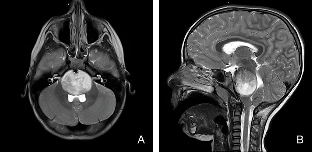
Ventriculomegaly is a condition in which the ventricles (fluid-filled spaces in the brain) are larger than usual. The brain has 4 ventricles – 2 at the top (on the left and right sides of the brain), one just below these two and one below the third one, near the top of the spine. Usually, the problem is with one or both of the top ventricles.