A colloid cyst is a non-cancerous mucus-containing growth in the brain. Colloid cysts are rare and are thought to have a congenital origin. There are typically no symptoms from a colloid cyst, but sometimes they can slowly expand in size, causing headaches, nausea/vomiting, or visual changes, particularly in people over the age of 30 years old.
Subarachnoid cisterns are enlarged pockets of cerebrospinal fluid (CSF) located between the two innermost brain layers (meninges). The quadrigeminal cistern is one of the subarachnoid cisterns. It is located at the posterior aspect of the midbrain and third ventricle. The quadrigeminal cistern also contains several blood vessels, nerves, and the pineal gland.
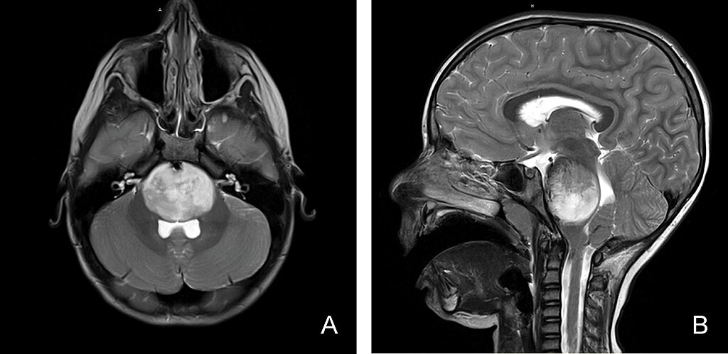
Areas with high water or protein content show up “bright” (hyperintense) on certain MRI sequences. There is a hyperintense spot (lesion) of the quadrigeminal cistern.
Intracranial lipomas are rare benign (non-cancerous) fat-containing lesions. They are congenital (present at birth) and account for 0.1 to 0.5% of all primary brain tumors. Quadrigeminal cistern lipomas are slow-growing lesions that makeup approximately 25% of intracranial lipomas. They are usually asymptomatic (do not cause symptoms), unless they are large enough to produce symptoms due to compression of nearby brain tissue or structures (mass effect). Symptoms, if present may include intracranial hypertension (a build-up of pressure around the brain), seizures, or hydrocephalus (a build-up of cerebrospinal fluid inside the brain).
A developmental venous anomaly (DVA), also known as venous angioma, is a congenital (present at birth) and benign (non-cancerous) malformation of the blood vessels. It is an irregular arrangement of small veins which drain into a larger central vein. They can occur anywhere in the body but are found most often in the brain or spinal cord.
DVAs are usually asymptomatic (without symptoms) and found incidentally (in passing when looking for something else). Sometimes DVAs can cause headaches; less commonly, DVAs can cause seizures, neurologic deficits and bleeding in the brain or spinal cord.
A harmless, congenital, thin-walled pocket of cerebrospinal fluid.
This is when extra CSF is surrounding one of the brain blood vessels. Enlarged perivascular spaces in the brain are common, but can sometimes be associated with cerebral small vessel disease.
The basilar artery is a blood vessel located at the back of the brain. It supplies oxygen-rich blood to portions of the brain and central nervous system. Ectasia of the basilar artery is a dilation of the artery that is not defined as an aneurysm (an abnormal bulge or dilation that occurs in the wall of the blood vessel). Ectasia of the basilar artery may be a marker for a high risk of stroke. Symptoms, if present, are related to compression of nearby cranial nerves or brain structures and may include headache and visual disturbances, including vision loss.