The pineal gland is located deep in the center of the brain, and is responsible for producing melatonin, a hormone which helps maintain our natural sleep-wake cycle. For unknown reasons, cysts (thin-walled fluid collections) can form within the pineal gland. The majority of pineal cysts stay stable in size or even shrink away. In rare cases, pineal cysts can grow over time and block the flow of cerebrospinal fluid (CSF), causing hydrocephalus (enlargement of the normal fluid cavities in the brain [ventricles]) and symptoms such as headache, nausea and vomiting, lethargy, confusion and double vision. If you develop these symptoms, it is a medical emergency requiring immediate care in a hospital.
Phthisis bulbi is a degenerative eye condition that is characterized by having severe irregular eye shrinkage and calcification (calcium buildup in the tissues of the eye that hardens over time). Common causes of phthisis bulbi include severe trauma to the eye, complications from eye surgery, inflammation, infection, and retinal detachment. Since this is a degenerative condition, symptoms such as changes in vision (e.g. blurred, cloudy vision), pain, and visual loss may worsen over time. Early treatment of the underlying cause is the best strategy to avoid and prevent complete vision loss.
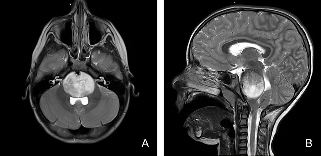
The pituitary gland is a small gland that sits in the sella turcica, a saddle-shaped depression located in the bone at the base of the skull. It produces hormones that control many different processes in the body, including metabolism, growth and reproduction. A cyst (fluid-filled pouch) can also arise from the pituitary gland. Pituitary cysts are not cancerous. Sometimes the fluid increases, putting pressure on the pituitary gland or on nearby nerves or brain tissue and causing symptoms such as headaches, visual impairment, and/or hormone changes.
The pituitary gland is a small gland that sits in the sella turcica, a saddle-shaped depression located in the bone at the base of the skull. It produces hormones that control many different processes in the body, including metabolism, growth and reproduction. Pituitary adenomas are the most common benign growth of the pituitary that may appear as a nodule, and sometimes hormone production can be affected. Pituitary adenomas can occur at any age, but are most common in people in their 30s or 40s. When an adenoma is less than 1 cm, it is called a microadenoma.
The pituitary gland is a small gland that sits in the sella turcica, a saddle-shaped depression located in the bone at the base of the skull. It produces hormones that control many different processes in the body, including metabolism, growth and reproduction. A nodule is a growth or lump in the body that could be benign (non-cancerous) or malignant (cancerous).
This is a common anatomic variant (estimated 10-30% of people) where the carotid artery (instead of the basilar artery) supplies blood to the posterior cerebral artery of the brain. This is a congenital (present from birth) condition, and no further follow-up or evaluation is needed for this finding.