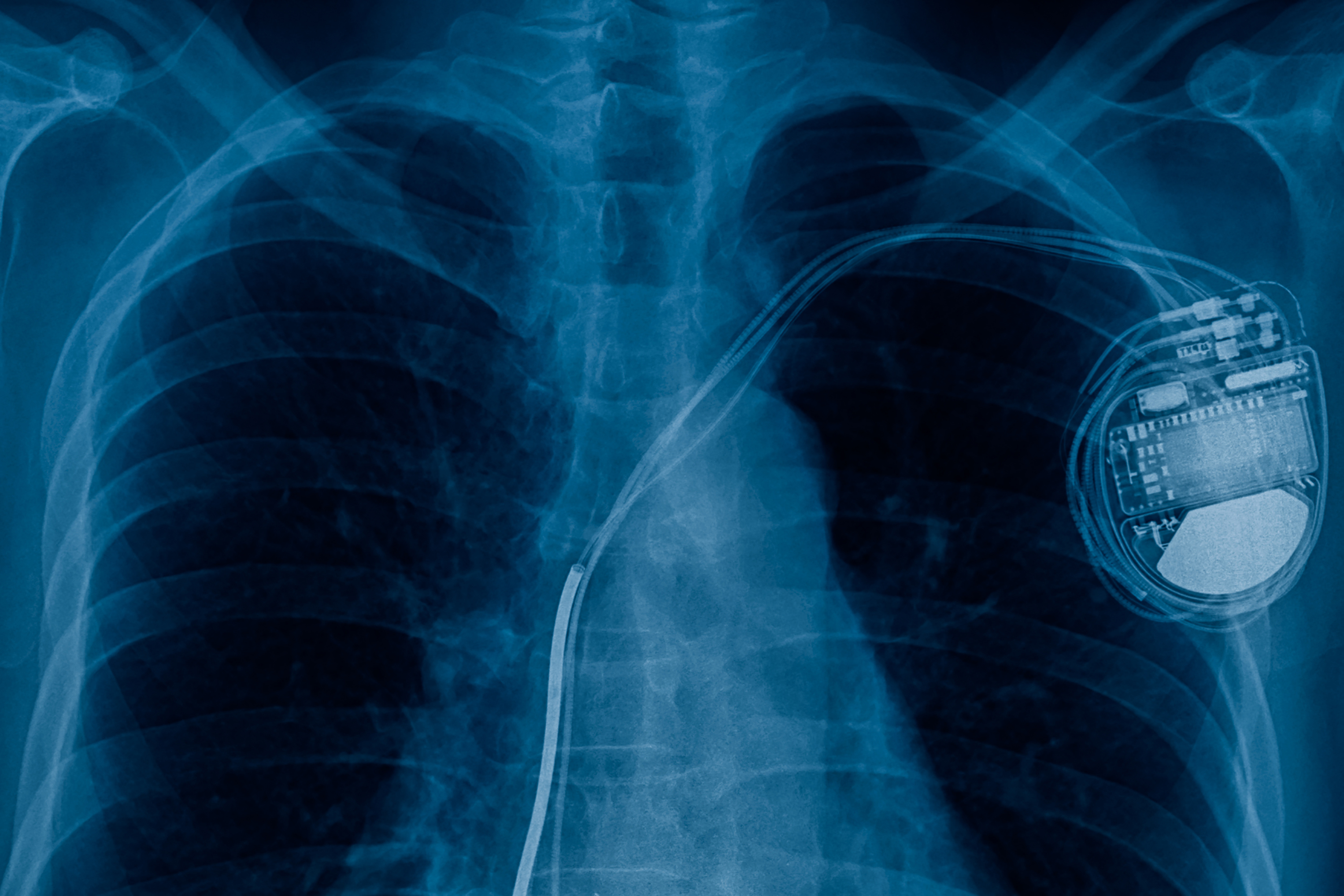
A pacemaker is a small, metal, electrical device that is inserted into the chest. It is used to treat arrhythmias (abnormal heart rhythms), for example, if the heart beats too slowly, known as bradycardia1.
Biventricular pacemakers help the chambers beat in sync and can treat heart failure2. Another type of metal cardiac device is an implantable cardioverter defibrillator (ICD), these are used to treat patients who have had, or are at risk of, life-threatening arrhythmias3.
The way they work is to deliver a shock to the heart and return it to a normal rhythm if it beats too quickly or abnormally4. Most of the time, patients with pacemakers or ICDs do not notice the device and live normal lives. Issues can arise, however, if they require an MRI scan.
As MRI scans use strong magnetic fields, they can disrupt and damage metal devices such as pacemakers5,6. Historically, having a pacemaker was a clear contraindication to having an MRI scan; however, newer devices are MRI compatible, and some research has shown that even older devices are safe.
What Happens During an MRI?
Magnetic resonance imaging (MRI) uses strong magnetic fields and radio waves to produce detailed internal images of the body7. It is a non-invasive imaging test and gives information on various structures, including organs, muscles, bones and blood vessels.
An MRI machine is a large, tube-shaped structure which can look like a narrow tunnel. Before the scan, your clinician will ask you to complete a safety questionnaire to check for any contraindications. If safe to proceed, the scan will be arranged. Read on to find out what happens during an MRI scan.
Step by Step:
- You will be asked to remove all clothing and given a gown. You will also need to remove all jewellery, glasses, hearing aids, removable dental work, hairpins or any other objects which may cause interference.
- If intravenous (IV) contrast is required, an intravenous line will be inserted. Your clinician will ask if you have any allergies prior to this. It is important to note that an Ezra scan does not require contrast.
- You will be asked to lie on a table and remain still. The table will move into the cylindrical scanner.
- The radiographer will be in the scanner control room. They will have a full view of you at all times and be in constant communication via a speaker.
- Earplugs or a headset will be provided to help block out the noise. You will hear a clicking sound as the scan is in progress.
- You may be instructed to hold your breath for very short intervals, depending on the area of your body which is being scanned. It will not be longer than a few seconds.
- If IV contrast is used, you will be warned that you may experience a metallic taste in your mouth, flushing sensation, coldness, itching, nausea or vomiting. This should only be for a brief moment. If they persist or you have any difficulty breathing, sweating or heart palpitations, then notify the technologist.
- Once complete, the table will move out of the scanner, and you will be helped off.
- An MRI scan is safe and should be pain-free; you may feel discomfort from having to lie flat and still for a length of time. Let the radiographer know if you have had a recent injury or procedure which may cause discomfort, so measures can be taken to help.
Understanding Pacemakers and MRI Scans
.jpeg)
As discussed, MRI scans use strong magnetic fields and radio waves to produce detailed internal images of the body. Unlike X-rays and CT scans, MRI scans do not use 8. That does not mean, however, that they come without risks.
The strong magnetic fields can reprogram or turn pacemakers off9. Subsequently, in patients with pacemakers or other implantable cardiac devices, MRI scans have been discouraged or prohibited, and alternative, often less effective, imaging techniques are used.
Due to an increasing number of individuals with pacemakers or ICDs, research has been carried out to better understand the risk associated with MRI scans. This is particularly relevant due to the increasing use of MRI scans in diagnostic medicine as well as for screening. Over half a million pacemakers have been implanted in the UK10.
There is growing evidence that MRI scans can be performed safely in patients with non-MR conditional pacemakers and ICDs11. A large UK registry study was conducted, including over 970 patients and more than 1,100 MRI scans of patients with both MR-conditional and non-MR-conditional pacemakers or ICDs, including higher-risk groups like pacemaker-dependent and ICD patients12. The study found no lead failures and only clinically insignificant changes in device parameters, showing comparable safety across MRI-conditional and non-MRI-conditional devices when appropriate protocols are followed.
A systematic review and meta-analysis also confirmed the safety of MRI scanning in patients with non-conditional cardiac implantable electronic devices (CIED)13. It must be noted, however, that a strict selection and monitoring protocol needs to be utilised. This involves device evaluation and peri-procedure programming, and close patient monitoring during the scan.
This requires experienced personnel as well as significant time investment. In many studies, the MRI procedure was supervised by cardiac technicians who are experienced in device programming, as well as cardiac nurses or a cardiologist who is trained in advanced cardiac life support.
In summary, for these scans to be performed safely in patients with CIEDs, there needs to be significant care and time taken to ensure the correct protocol is followed. For Ezra, patient safety is an absolute priority, and we will refer you to discuss any potential interference from metal objects such as pacemakers with your MRI technologist.
Producing MRI-compatible devices without the need for interrogation, monitoring, or programming is the ultimate goal, with the hope that patients with cardiac devices can be assured of their safety during an MRI scan in the future.
Other Options for Scans with Pacemaker
Depending on the part of your body which needs to be scanned, there are alternative options to an MRI.
CT scans are often used; these use ionising radiation instead of magnetic fields, and therefore, patients with metal devices are safe. CT scans create images of bones and soft tissues; however, they are not as effective as an MRI at differentiating subtle differences between soft tissues. Learn more about tests and imaging techniques which are used to test for cancer.
Ultrasound is also an imaging modality which is safe for patients with pacemakers. Ultimately, it is up to the treating physician to decide which scan is most appropriate in the context of the clinical picture.
It is important to discuss the issue with a clinician before proceeding with any scan. Such decisions are made on a risk/benefit basis. The benefit of an MRI may outweigh the perceived risks if it can be performed under the correct protocol.
MRI and Pacemaker: Summary
Historically, patients with a pacemaker were not able to get MRI scans due to the risk of damaging the device or surrounding tissue. Fortunately, due to evolving technology, most new pacemakers and ICDs are now MRI compatible.
Many individuals, however, have older devices that are deemed non-MR conditional. There has been promising evidence which demonstrates that MRI scans can be performed safely in patients with these devices, but strict protocols and monitoring are required.
If an MRI is recommended and you have a pacemaker or ICD, your GP must be aware so they can assess if it is a compatible device. Additionally, if you have had one recently inserted, you will need to wait a period of time prior to the scan. Be sure to consult your doctor.
Ezra does not currently recommend a multi-region MRI for individuals with pacemakers. For those without such devices, however, it is a safe, non-invasive imaging modality which gives information on over 14 different organs. Changes could be detected and treated before they become symptomatic or problematic, so be proactive about your health and consider booking a scan today.
Understand your risk for cancer with our 5 minute quiz.
Our scan is designed to detect potential cancer early.
References
1. Pacemakers. British Heart Foundation. Accessed October 17, 2025. https://www.bhf.org.uk/informationsupport/treatments/pacemakers
2. Pacemakers - How They Work | NHLBI, NIH. March 24, 2022. Accessed October 17, 2025. https://www.nhlbi.nih.gov/health/pacemakers/how-it-works
3. Implantable cardioverter defibrillator (ICD). British Heart Foundation. Accessed October 17, 2025. https://www.bhf.org.uk/informationsupport/treatments/implantable-cardioverter-defibrillator
4. Pacemaker implantation. nhs.uk. October 20, 2017. Accessed October 17, 2025. https://www.nhs.uk/tests-and-treatments/pacemaker-implantation/
5. Why might I not be able to have an MRI scan? British Heart Foundation. Accessed October 17, 2025. https://www.bhf.org.uk/informationsupport/heart-matters-magazine/medical/ask-the-experts/pacemaker-and-mri-scan
6. Bovenschulte H, Schlüter-Brust K, Liebig T, Erdmann E, Eysel P, Zobel C. MRI in Patients With Pacemakers. Dtsch Arztebl Int. 2012;109(15):270-275. doi:10.3238/arztebl.2012.0270
7. MRI scan. nhs.uk. October 23, 2017. Accessed October 17, 2025. https://www.nhs.uk/tests-and-treatments/mri-scan/
8. Magnetic Resonance Imaging (MRI). National Institute of Biomedical Imaging and Bioengineering. Accessed October 17, 2025. https://www.nibib.nih.gov/science-education/science-topics/magnetic-resonance-imaging-mri
9. Potential Hazards and Risks. UCSF Radiology. January 20, 2016. Accessed October 17, 2025. https://radiology.ucsf.edu/patient-care/patient-safety/mri/potential-hazards-risks
10. From the procedure to recovery: Frequently asked pacemaker questions. British Heart Foundation. Accessed October 17, 2025. https://www.bhf.org.uk/informationsupport/heart-matters-magazine/medical/how-does-a-pacemaker-work/frequently-asked-pacemaker-questionse
11. Bhuva A, Charles-Edwards G, Ashmore J, et al. Joint British Society consensus recommendations for magnetic resonance imaging for patients with cardiac implantable electronic devices. Heart. 2024;110(4):e3-e3. doi:10.1136/heartjnl-2022-320810
12. Bhuva AN, Moralee R, Brunker T, et al. Evidence to support magnetic resonance conditional labelling of all pacemaker and defibrillator leads in patients with cardiac implantable electronic devices. Eur Heart J. 2021;43(26):2469-2478. doi:10.1093/eurheartj/ehab350
13. Munawar DA, Chan JEZ, Emami M, et al. Magnetic resonance imaging in non-conditional pacemakers and implantable cardioverter-defibrillators: a systematic review and meta-analysis. Europace. 2020;22(2):288-298. doi:10.1093/europace/euz343


