
Key Takeaways:
- Magnetic resonance imaging has been around for over a quarter of a century.
- Nuclear magnetic resonance (NMR) is the basis of medical MRIs.
- In 1969, Dr. Raymond Damadian hypothesized and demonstrated that magnetic resonance could differentiate cancer cells from non-cancerous cells.
- Technology advances have significantly reduced the time required for individual MR slices.
The history of MRIs goes back to the 1930s, when researchers, scientists, and doctors first developed ever-improving magnetic resonance imaging (MRI) scans.
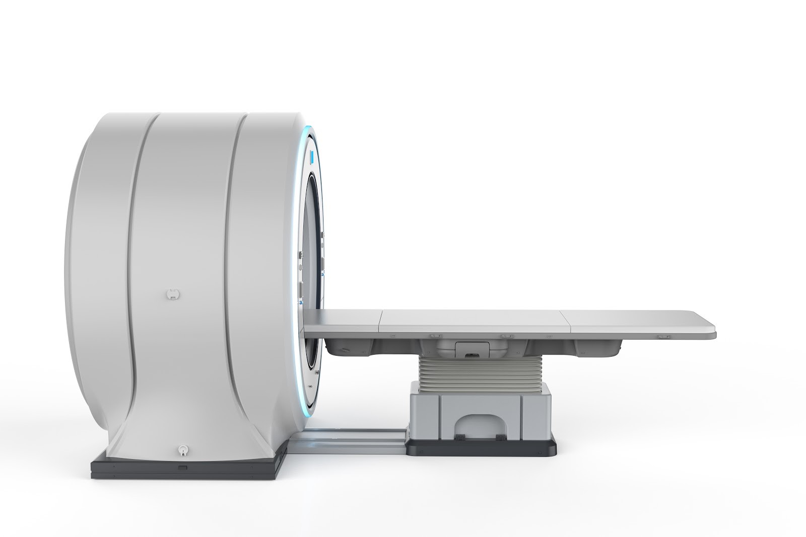
Radiologists use magnetic resonance (MR) imaging, which uses radio waves in a strong magnetic field to produce soft and bony tissue images to help doctors detect cancer and other diseases.
The Development of Magnetic Resonance Imaging (MRI Scans)
The development of MRI technology depended on many individuals’ research throughout most of the 20th century and into the 21st century. Notables include physicists Sir Peter Mansfield, I.I. Rabi, Edward Purcell, and Felix Bloch.
Chemists involved in magnetic resonance imaging research include researchers Paul Lauterbur, Erik Odeblad, Gunnar Lindström, and Dr. Raymond Damadian.
Today, MRI technology continues to advance as medical imaging becomes more important in cancer prevention and detection as well as medical diagnosis for cancer and other conditions.
MRI technology can help doctors arrive at a medical diagnosis by differentiating between healthy tissue and cancerous cells.
The History of MRI Technology
The history of MRI technology begins with the study of magnetic resonance or how electrons’ and atoms’ nuclei respond to magnetism.
In the 1930s, physicist I.I. Rabi developed a way to measure the magnetic properties (spin) and sodium movement. In his work, Rabi developed a form of MR imaging called nuclear magnetic resonance (NMR). That work became the basis of medical MRIs.
During the 1940s, physicists Felix Bloch and Edward Purcell, working independently, studied the atomic and molecular magnetic resonance properties of solids and liquids. Their research later allowed MRI scanners to use the body’s water content to develop magnetic resonance images.
In 1952, Purcel and Bloch won the Nobel Prize in physics. Publishing their paper in 1955, Gunnar Lindström and Erik Odebladalso, two Swedish researchers, also investigated NMR’s uses. They theorized that the differences in water and biological tissue response were because distinct tissues absorb and organize water molecules differently.
In 1969, Dr. Raymond Damadian hypothesized that magnetic resonance could differentiate cancer cells from non-cancerous cells, and he successfully demonstrated his hypothesis with rats. Damadian found that different kinds of tissue emissions had different response lengths, with longer response signals from cancerous tissue.
Damadian, a physician, scientist, and professor at the Downstate Medical Center State University of New York in Brooklyn, developed the original idea of applying NMR to medical imaging (MRI). His research into the sodium and potassium content of living cells led him to his first experiments with nuclear magnetic resonance (NMR).
Damadian discovered that it’s possible to distinguish between cancer cells and healthy tissue through nuclear magnetic resonance (NMR) because of the differences in relaxation times between the two. From that research, he proposed an MR body scanner in 1969.
In 1972, Damadian filed the first patent of MRI technology. Upon approval of his patent in 1974, Damadian designed and built a full-body MRI machine. The first MR (magnetic resonance) scanning machine, which he called “Indomitable,” is now in the Smithsonian.
On July 3, 1977, Damadian achieved the first human NMR image — a cross-section of his postgraduate assistant Larry Minkoff’s chest. The image revealed Minkoff’s heart, lungs, vertebrae, and musculature and became the method known as magnetic resonance imaging (MRI).
The First Commercial MRI Machine
In 1978, Damadian founded FONAR Corporation (field focused nuclear magnetic resonance). FONAR produced the first commercially available MRI machine in 1980.
The corporation currently has 15 MRI scanning centers in the U.S. In 1985, FONAR introduced the first mobile MRI, often used in the ICU where it may be a danger to move the patient, or in an ambulance or emergency disaster setting.
In 2007, The Intellectual Properties Owners Association Education Foundation recognized FONAR’s Upright Multi-Positional MRI as The Invention of the Year. Collaborating with Wilson Greatbatch, Damadian also developed an MRI-compatible pacemaker.
From 1972 through 1980, chemist Paul C. Lauterbur began developing magnetic resonance imaging methods. He reconstructed two-dimensional images using magnetic field gradients by stacking the images to create 3D images.
Lauterbur also developed a technique, which he called zeugmatography.
Zeugmatography joins spatially defined radiofrequency field gradients and a magnetic field. In that way, Lauterbur could generate a 2-D display of tissues’ proton density and relaxation times. Lauterbur realized that using a non-uniform magnetic field allowed the precise localization of structures.
At the same time, physicist Sir Peter Mansfield began work to reduce the time required for complete magnetic resonance scans. Using a new technique, Mansfield took images of a finger in 15-23 minutes. This was the first time a human body part was successfully scanned with NMR technology.
Technology advances in gradient and digital data acquisition have reduced the time required for individual MR slices to 50-100 msec.
Known as echo-planar imaging (EPI), the shortened time frame minimizes the errors caused by patient movement. Using EPI, Mansfield’s team produced the first biologic EPI images of a beating rabbit heart. Later they created images of an infant human heart in 1981 and again in 1983.
Working independently and building on Damadian’s ideas, Lauterbur and Mansfield shared the Nobel Prize in Physiology or Medicine in 2003 for their magnetic resonance imaging discoveries.
MRSI combines the MRI imaging capabilities with the analytical capabilities of nuclear magnetic resonance (NMR) spectroscopy, allowing metabolite distribution monitoring within the body.
Radiology

Radiology specialists use radiation to detect, diagnose, and treat disease. Radiology includes ionizing radiation, such as X-rays and computed tomography, and non-ionizing radiation, such as ultrasounds.
Radiologists interpret the resulting images to diagnose conditions and diseases. Radiology also assists physicians and surgeons in treating, locating, and defining the area of concern.
Computed Tomography (CT) Scan
Sir Godfrey Newbold Hounsfield CBE, FRS., was an English electrical engineer who developed X-ray computed tomography.
The Hounsfield Scale, which measures radiodensity in CT scans, is named for him. Also, Hounsfield shared the 1979 Nobel Prize for Physiology or Medicine with Allan McLeod Cormack for his work.
MRI Scan
Rapid advances in MR imaging have highlighted radiology as a focal point in modern academic medicine. Mammography, cardiology, sports injuries, and neurology use MR images, contributing to improved patient outcomes.
Magnetic Resonance (MR) spectroscopy is a non-intrusive diagnostic method to measure biochemical changes in the brain, especially when tumors are present.
Using a conventional MRI, MR spectroscopy compares the chemical compositions of normal brain tissue and abnormal tumor tissue. MR spectroscopy analyzes such molecules as hydrogen ions or protons. Proton spectroscopy is the most commonly used form of MR spectroscopy.
Diffusion MRI is another magnetic resonance imaging technique that uses a contrast mechanism determined by water molecules’ microscopic mobility.
In living patients, tissue structures, such as cell membranes, slow water molecules’ diffusive motion. The diffusion distinguishes between pure fluid and densely packed cells.
MRI scanners are now used worldwide in a wide range of medical facilities. Healthcare providers order and perform millions of MRI scans each year to screen patients for cancer, identify tissue injuries and organ dysfunction, and monitor treatment effectiveness.
Because MRI scans use magnets rather than radiation, they are one of the safest imaging modalities available.
Take Control of Your Health by Getting an Elective Full-Body MRI Screening
At ezra, we believe the best defense against cancer is early detection. It’s easy to schedule a regular annual full-body MRI scan and take control of your own health and wellbeing.
Curious about your susceptibility to cancer?
Take our five-minute quiz to understand your risk level. With just a bit of information about your family background, your medical history, and your lifestyle, we can quickly give you a snapshot of your risk scores.


