Magnetic resonance imaging (MRI) is an essential part of modern medicine. MRIs provide detailed images of internal body structures without using harmful ionizing radiation. They are particularly useful for detecting different types of cancer and determining how far the disease has progressed. A specific MRI scan called brain and orbit MRI creates detailed pictures of the brain and eye sockets (orbits). Clinicians use these scans to detect tumors, strokes, infections, and neurological diseases. This article will explain what brain and orbit MRI scans are, what to expect during the procedure, their medical applications, and future developments of this technique in neurology and ophthalmology.
Understanding Brain and Orbit MRI Scans: What You Need to Know
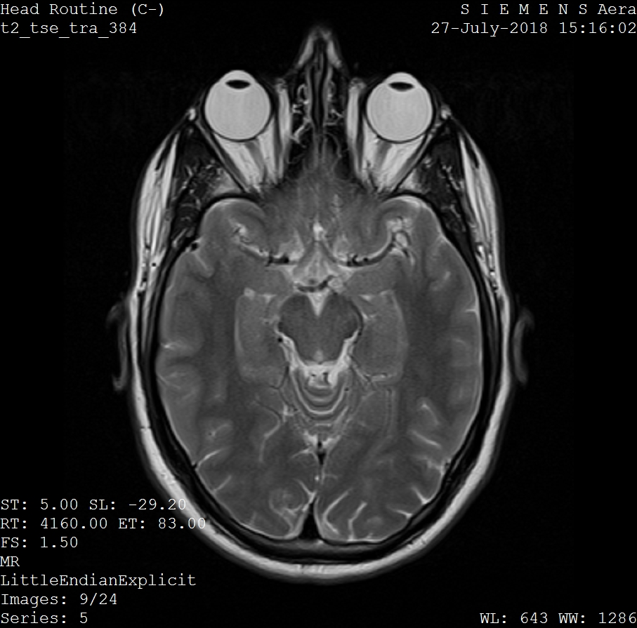
What Are Brain and Orbit MRI Scans?
Brain and orbit MRI scans are specialized imaging procedures that use magnetic fields and radio waves to create detailed pictures of the brain, eye sockets, and surrounding structures without using radiation.
Basics of MRI Technology
MRIs use a combination of a strong magnetic field and radio waves to generate highly detailed images of internal body structures. During an MRI, the magnetic field causes the hydrogen protons in the body to align (like a compass needle pointing north). When the radio waves are switched on, the protons are forced out of alignment. When the radio waves are switched off, the protons realign and emit signals that create the image.
Understanding the Brain and Orbit Regions
You already know what the brain is, but the term “orbital” is less common. The orbitals are the sockets in the skull that hold the eyes. They also contain the nerves, blood vessels, muscles, and other tissues that allow the eyes to function. The brain and orbitals are close together, making it easier to scan both simultaneously compared to more distant body sites. Healthcare providers can choose to image different areas of the brain and the orbitals depending on the disease they are examining.
Purpose of Brain and Orbit MRI Scans
These scans help assess, diagnose, and monitor a variety of health issues related to the eyes and neurological tissue. Radiologists use different settings on MRI machines to scan for abnormalities that include but are not limited to, cancers such as retinoblastoma and glioma, multiple sclerosis, stroke, microbial infections, retinal detachment, endophthalmitis (inflammation of the inner eye coating), neuropathies, and posttraumatic hemorrhagic cysts.
The Procedure: What to Expect During an MRI Scan
Preparing for the MRI
Before an MRI, a healthcare professional will talk you through the procedure and relevant safety precautions. You will be asked to remove metal objects and disclose the presence of metal objects within your body, such as pacemakers. If you are prone to claustrophobia, you should tell your healthcare provider ahead of time so they can help alleviate your concerns and make the procedure as comfortable for you as possible. Remember that MRIs are not painful and are non-invasive.
The MRI Scan Process
You will be asked to remove your clothing, put on a gown, and lay down on the MRI table. You will likely be provided with a support cushion to help keep your eyes and head steady during the scan. MRI machines make loud noises as they take images, so you will be provided with earplugs or earmuffs to protect you from this noise. The procedure typically takes 30-45 minutes. If you need to move or take a break during the procedure, it’s important to tell your technologist beforehand. They will communicate with you via an intercom.
Post-Scan Procedures and Results
MRI is a non-invasive procedure, which means you will be allowed to leave and go about your day as usual after the scan is complete. Trained radiologists will study the images to determine the presence of abnormalities and it typically takes one to two weeks to receive results. This time scale may be shorter or longer depending on factors such as backlogs and the urgency of your scan.
Innovations in Brain and Orbit MRI Scans
Improvements in brain and orbit MRI technology mean doctors can more accurately assess your health and make better decisions.
Advancements in MRI Technology
Dynamic contrast-enhanced MRI uses a contrast agent and takes multiple images in rapid succession to achieve better images of tiny blood vessels. Studies have shown that this technique is better at determining whether lesions in the eye are benign or malignant. Dynamic MR angiography is another innovation that allows doctors to assess how well blood is flowing in the tissue being imaged. This is important as abnormal blood flow can indicate the presence of inherited malformations in blood vessels.
AI Integration in MRI Analysis
AI is beginning to play a massive role in MRI procedures. It can be used to assess images more quickly and accurately and find abnormalities that are more difficult to detect with the naked eye. Deep learning algorithms can process large amounts of scan data, which means healthcare professionals get results more quickly and can direct appropriate patient care more efficiently.
Future of MRI in Neurology and Ophthalmology
MRI machines are being designed with stronger magnetic fields, significantly increasing image resolution. Currently, MRIs with ultra-high magnetic fields use 7 Tesla. For context, conventional MRI machines use 1.5 to 3 Tesla, and the world's most powerful MRI machine uses 11.7 Tesla. Higher resolution means tiny lesions and other abnormalities can be detected earlier, and clinicians and researchers can gain deeper insights into a wide range of diseases. Multimodal optic nerve imaging (including MRI) is also being advanced to improve the diagnosis of optical neuropathies.
Applications and Benefits of Brain and Orbit MRI Scans
At the start of the article, we discussed how hydrogen protons emit signals that produce images during MRIs. This works because these protons behave differently depending on what tissue they are part of, allowing clinicians to tell the difference between normal tissues and ones that indicate the presence of disease.
Diagnosing Neurological Conditions
Brain and orbit MRI scans are used to diagnose conditions like tumors, strokes, multiple sclerosis, and other neurological issues. Myelin is a fatty tissue that acts as a protective layer around nerve fibers. In multiple sclerosis, this myelin layer is gradually lost. The water content of fat is different from that of other tissue types, which means that fatty tissue (like myelin) gives a different signal than surrounding tissues during MRI scans. This means that MRI scans can detect where myelin has been lost and help diagnose multiple sclerosis.
Detecting Orbit and Eye Conditions
MRI helps identify conditions affecting the eyes and surrounding structures, such as orbital tumors or optic nerve issues. For example, retinoblastomas grow inside the vitreous chamber (the largest chamber of the eye), and MRI scans can identify retinoblastomas because they are denser than the fluid within the vitreous chamber. Optic nerve gliomas are clearly visible on MRI as enlargements of the optic nerve and are easily comparable to the unaffected nerve on the other side.
Non-Invasive Imaging Benefits
MRIs provide non-invasive imaging that does not use ionizing radiation like other imaging techniques like computed tomography and X-ray. It is a significantly safer technique than these alternatives and is suitable for repeated use, which benefits individuals for whom regular scans are recommended. It is a painless procedure, and while it may trigger claustrophobia in some individuals, technologies like open MRI machines are available to counteract this.

Conclusion
Brain and orbit MRI scans are crucial for diagnosing many health issues affecting the eyes and nervous system. Advances in MRI technologies, including the use of AI, mean that clinicians can provide more timely and accurate healthcare to patients, which can make a huge difference in patient outcomes. MRIs are safe, painless, and efficient. They usually take less than an hour to complete and allow patients to get on with their day immediately afterward. It’s normal to have concerns about medical scans, so be sure to discuss any concerns you have about MRIs with your healthcare provider.
If you want to be proactive about your health, why not book an Ezra full body MRI? Our annual scan catches potential cancer earlier, leveraging AI through the screening process to make it more efficient, affordable and faster.


