Ovarian cancer is the fifth most common cause of cancer-related death in women in the US and the eighth most common globally. There are two broad types of ovarian cancer, epithelial and non-epithelial, accounting for 95 and 5 percent of cases, respectively. A primary challenge when dealing with ovarian cancer is that it does not present with obvious or specific symptoms at the early stages.
This means that when women develop symptoms like post-menopausal bleeding and abdominal pain and go for screening, the disease is already likely to have progressed to a later stage, which is less treatable and has a worse survival rate. Another issue is the lack of effective screening tools to catch ovarian cancer at the early stages.
Magnetic resonance imaging (MRI) is a central component of screening programs for many types of cancer, though it is not considered a primary method for detecting ovarian cancers. Despite this, MRI can be used to detect ovarian cancer in some instances, though it has some limitations that reduce its utility. This article will explore the accuracy of MRI (Magnetic Resonance Imaging) in detecting ovarian cancer and the technology behind how it works.
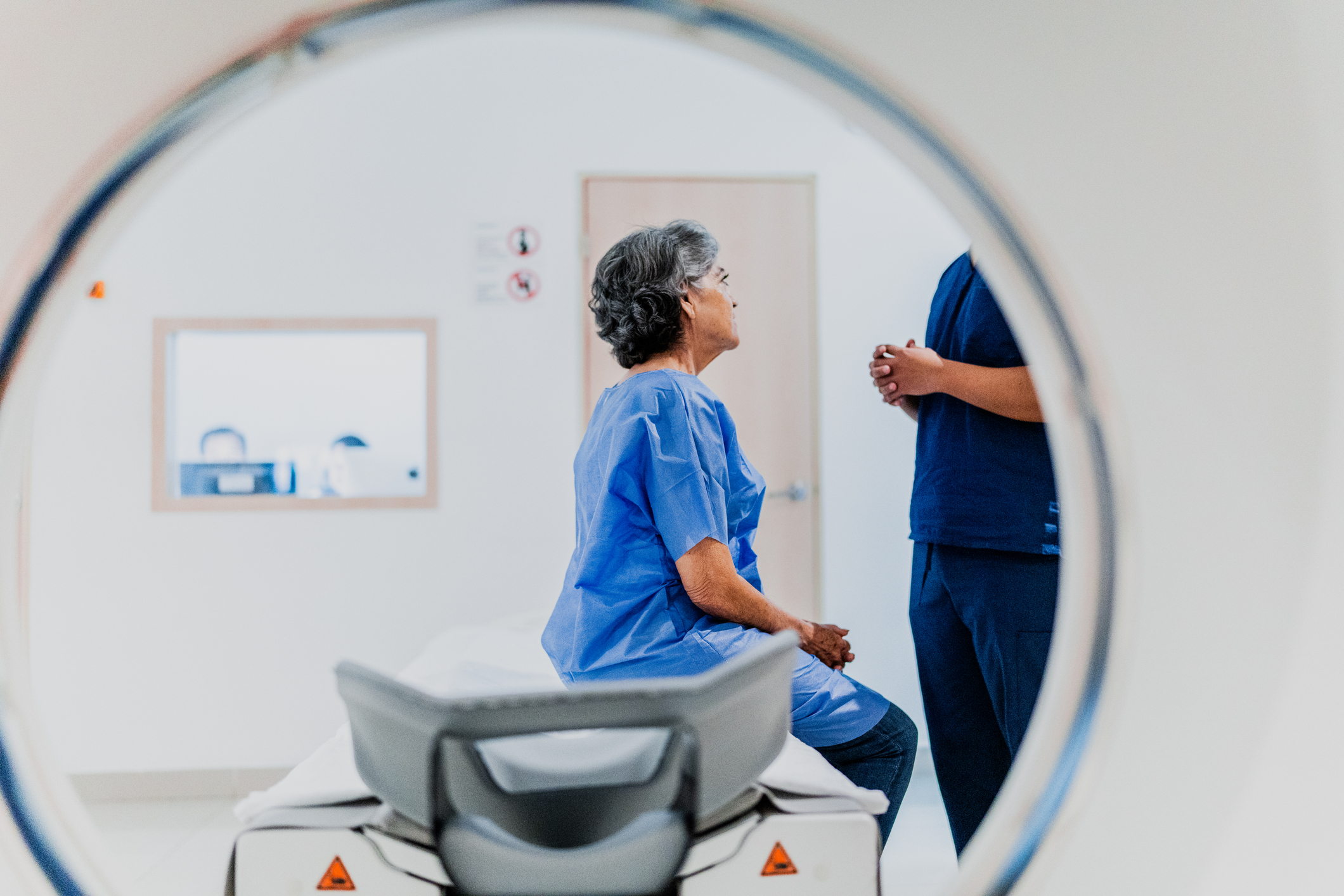
What is an MRI?
MRI is a noninvasive medical imaging technique used to diagnose a variety of conditions. It does not use harmful radiation, giving it an advantage over other imaging techniques like X-rays.
How MRI Works
MRI uses a strong magnetic field and radio waves to generate detailed images of the body's internal structures. Specifically, protons in the body's water molecules align with the magnetic field, and when radio waves are applied, they emit signals that are captured and transformed into images by a computer.
Advancements in MRI technology include the use of more powerful magnetic fields to generate higher-quality images. These days, many machines use 3 teslas (3T) of magnetic force, compared to 0.5 and 1.5 that were used previously. Artificial intelligence (AI) is becoming increasingly crucial in MRI technology, enabling faster image reconstruction and data analysis.
MRI Techniques for Ovarian Cancer Detection
MRI pulse sequences are sets of instructions for the MRI machine, which determine how it will use magnetic fields and radio waves to create detailed images of the body. Different sequences, like T1-weighted or T2-weighted, highlight various tissues or abnormalities for diagnostic purposes.
T1-weighted: This technique makes fat appear bright, and fluids appear dark, which helps in assessing anatomy and detecting abnormalities like tumors.
T2-weighted: This technique highlights fluid-filled areas, making them appear brighter. It is useful for identifying inflammation, edema (swelling caused by trapped fluid), and other fluid-related abnormalities.
In practice, T1- and T2-weighted approaches are used simultaneously to detect abnormalities like tumors, which have different signal intensities compared to the surrounding healthy tissues.
Diffusion-weighted imaging (DWI): This MRI technique maps the movement of water molecules in tissues, helping to detect abnormalities like tumors by highlighting areas where water diffusion is restricted.
Comparing MRI with Other Imaging Techniques
Ultrasound, specifically transvaginal sonography, is generally the first imaging technique used for detecting ovarian cancer. Computed tomography (CT) and MRI techniques can also detect ovarian cancer, though they are more useful in determining the stage of the disease. Importantly, a recent study found that MRI outperformed CT in diagnosing ovarian cancer. MRI has also shown promise in detecting metastases that are more challenging to visualize with other techniques.
Diagnostic Accuracy of MRI in Ovarian Cancer
Diagnostic accuracy is essential for avoiding the risks associated with false positive and false negative results. This is particularly important in ovarian cancer, which often presents with nonspecific symptoms and is typically diagnosed at an advanced stage, making accurate and early detection critical for effective treatment and improved survival rates.
Sensitivity and Specificity
Sensitivity in diagnostic imaging is how well an MRI can correctly identify people who have an ovarian tumor. Specificity is how well it can correctly identify people who do not have a tumor. Both are crucial for accurate MRI results, reducing false positives and negatives. MRI has respective sensitivity and specificities of 96 and 91 percent for distinguishing malignant ovarian tumors from benign ones. Despite these statistics, MRI scans have been reported to struggle with distinguishing borderline cases of malignancy.
Distinguishing Between Benign and Malignant Masses
MRI allows for the evaluation of multiple tumor parameters that help to distinguish between benign and malignant tissue. For example, the presence of peritoneal nodules indicates an advanced stage of disease that has metastasized. The nature of the fluid found within an ovarian cyst helps differentiate between “almost certainly benign” and “low risk” masses, and MRI can assess this. Dark T2 or DWI staining of an abnormal lesion also indicates the mass is “almost certainly benign”. The presence of solid tissue indicates the presence of a malignant tumor, whose risk can be characterized by further examination using MRI.
Role of MRI in Ovarian Cancer Screening
Effective screening remains a significant challenge in ovarian cancer, and there is currently no standardized strategy for screening. In the absence of guidelines, many different imaging and diagnostic strategies are implemented depending on patient and tumor characteristics.
Limitations and Challenges
Serum cancer antigen (CA125) is a protein often found in higher levels in the blood of people with certain types of cancers, most notably ovarian cancer. Blood biomarkers like CA125 are incredibly important because they provide valuable diagnostic information, allowing for the early detection and monitoring of cancers without the need for invasive procedures to obtain a tumor biopsy. However, so far, clinical trials have failed to show a reduction in mortality of ovarian cancer with screening using CA125. However, in a recent study of women with a high risk of ovarian cancer due to genetic predisposition, regular MRI screening reduced mortality, suggesting a potential role for MRI in screening high-risk populations.
Integrating MRI into Diagnostic Pathways
While MRI is typically not the first imaging method used to diagnose ovarian cancer in patients presenting with symptoms, it can be used later on for cancer staging. For example, it is useful for detecting the invasion of tumor cells into the surrounding tissues and detecting metastases in distant organs like the brain. These factors are critical in diagnosing late-stage ovarian cancer and choosing the appropriate management plan.
Future Directions and Innovations
In addition to the advances in MRI technology described above, DCE (Dynamic Contrast)-MRI also shows promise in improving the accuracy of MRI in characterizing complex ovarian tumors. This type of MRI uses a special dye to highlight blood flow and tissue characteristics, helping doctors detect and analyze tumors.
Summary: How Accurate Is MRI in Detecting Ovarian Cancer
MRI plays a crucial role in detecting and diagnosing ovarian cancer, offering high sensitivity and specificity to distinguish between benign and malignant masses. MRI techniques, such as T1-weighted, T2-weighted, and diffusion-weighted imaging, provide detailed insights into tumor anatomy, characteristics, and disease staging. MRI is valuable in distinguishing between benign and malignant tissue despite its limitations in some borderline cases. It is especially useful in situations where other imaging methods are insufficient to grade tumors accurately.
While not typically the first-line imaging technique, MRI can characterize complex or ambiguous tumors, detect metastases, and evaluate invasion. Understanding its strengths and limitations is essential for healthcare professionals. These features make MRI a valuable tool to incorporate into the diagnostic pathway for ovarian cancer.
If you want to assess your risk for ovarian cancer, start with Ezra’s risk factor calculator. It’s easy to use and completely free and takes into consideration family history and genetic mutations like BRCA1 and previous cancer diagnosis. Alternatively, if you want to be proactive about your health status before any symptoms arise, consider booking an Ezra Full Body MRI. This painless, non-invasive scan can screen for potential cancer in up to 13 organs as well as 500 other conditions.


