
Mediastinal tumors are abnormally growing tissues within the chest cavity between the lungs, called the mediastinum. These tumors compress important organs like the heart, esophagus, and trachea, causing serious health issues. That’s why detecting them early and monitoring their growth is important to prevent damage to these vital organs. This blog will outline the growth rate of these tumors, what symptoms to watch out for, and why early detection could be life-saving.
What Is a Mediastinal Mass?
While “mediastinal mass” and “mediastinal tumor” both involve growths found in the mediastinum, they don’t necessarily mean the same thing. A mediastinal mass is a much broader term and usually describes a non-cancerous (benign) growth, like benign teratomas or cysts, while a mediastinal tumor means a cancerous (malignant) growth1. Many mediastinal masses are non-cancerous, but they may still compress vital organs, causing serious health concerns2.
Common types include thymomas, lymphomas, and germ cell masses, usually found in the anterior (front) mediastinum. In some people, a warning sign of cancer may include mediastinal adenopathy (swollen lymph nodes in the mediastinum), so keeping an eye on these warning signs can help with early detection3.
Location and Function of the Mediastinum
The mediastinum is found in the middle of the chest between the sternum (breastbone) and spine1. Tumors that develop in the mediastinum are dangerous because they can put pressure on vital organs like the heart, esophagus, and trachea, which are all essential for a healthy and functioning body1.
Common Types of Mediastinal Tumors
Thymoma - Typically slow growing and develops in the thymus gland, an essential organ in the immune system. This gland produces white blood cells and is critical for the body to fight infections and disease4.
Lymphoma - Typically fast growing, and develops in the lymphatic system, a critical part of the immune system’s defence. Examples of lymphomas include Hodgkin lymphoma and non-Hodgkin lymphoma5.
Germ cell tumors - Develop from germ cells (cells which make sperm and eggs) growing outside the ovaries and testes. They can be categorized into seminomas, which tend to be slower growing, and non-seminomas, which are faster growing5.
Neurogenic tumors - Develop from nerve tissue and are one of the most common mediastinal tumors found in the posterior (back) mediastinum6.
Is Mediastinal Adenopathy Cancer?
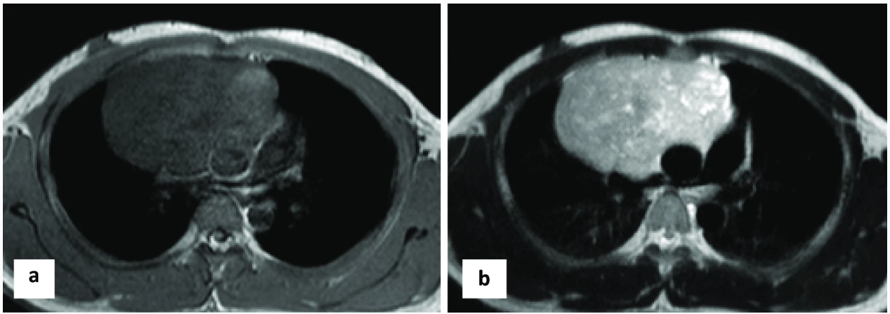
Mediastinal adenopathy means the swelling of lymph nodes in the chest. It can be a sign of infection, like upper respiratory infections, or inflammatory conditions. But it could also be a red flag for cancer, like Hodgkin and non-Hodgkin lymphoma3. When signs of mediastinal adenopathy are present, it’s important to talk to healthcare providers. They may recommend carrying out imaging tests, like a chest X-ray, computer tomography (CT) or magnetic resonance imaging (MRI) scan, which can help detect abnormal tissues1.
Mediastinal Cancer Symptoms
Respiratory and Chest Symptoms
Symptoms of a mediastinal tumor are usually due to important organs like the heart, lung, and airway being compressed, they can include1:
- Trouble swallowing
- Cough
- Wheezing
- Shortness of breath
- Hoarseness
- Coughing up blood
Systemic Symptoms
Some people also experience systemic symptoms like fatigue, unexplained weight loss, fever, chills, and night sweats1.
When to Seek Medical Advice
Early screening is the best way to detect abnormalities, especially if symptoms persist or worsen. Mediastinal tumors may remain silent as they develop, not producing any symptoms. As a result, they’re usually discovered at a late stage, and even accidentally after imaging tests for an unrelated health issue1.
How Fast Do Mediastinal Tumors Grow?
Mediastinal tumors grow at different rates depending on the type of tumor. For example, lymphomas typically grow fast, requiring urgent medical attention for the best possible outcome, whereas thymomas tend to grow more slowly, but still need careful monitoring5. Imaging techniques, such as CT and MRI scans, are essential in detecting abnormalities and monitoring how well the tumor responds to treatment.
Role of Imaging in Monitoring Growth
Imaging techniques like CT and MRI scans are great tools for detecting abnormalities, such as mediastinal masses and tumors1. They can help determine the tumor’s size, shape, and whether it has spread.
Importance of Biopsy
If imaging detects abnormalities, a biopsy of the tissue (mediastinoscopy) is typically the next step to check for cancer and guide treatment decisions.
Factors That Influence Tumor Growth
Tumor type - Slow-growing mediastinal tumors include thymomas, whereas aggressive tumors like lymphomas and non-seminoma germ cell tumors tend to grow faster5
Immune response - Inflammation from a collection of immune cells in the mediastinum has been linked to increased tumor growth7
Location - Anterior (front) and posterior (back) mediastinal tumors grow at varying rates, depending on the type of mediastinal tumor and areas of the body they impact1
Stage - Early-stage tumors often grow slowly and stay in one area of the body, while late-stage tumors are generally more advanced, grow faster, and may spread to other parts of the body8
Diagnosis & Stages
Imaging tools like chest X-ray, CT, MRI, and positron emission tomography (PET) scans help detect mediastinal masses and tumors. The next step is usually a biopsy to confirm cancer and determine its stage (I-IV), based on the location of the primary tumor, whether it has spread to nearby lymph nodes, and whether it has spread to distant parts of the body8. Early-stage tumors may be treated by surgery, whereas advanced tumors (stage III-IV) often require radiation therapy and/or chemotherapy.
Stage I - Tumor is in the mediastinum with no spread to other parts of the body
Stage II - Mediastinal metastasis (spread of tumor) to surrounding tissues or lymph nodes within the mediastinum
Stage III - More extensive spread to nearby organs such as the lungs, heart, or major blood vessels
Stage VI - Cancer has spread to distant parts of the body, such as the bones or liver
Mediastinal Metastasis
Sometimes mediastinal tumors can spread to other parts of the body, which is called metastasis. The sites for metastasis usually depend on the type of mediastinal tumor; for example, germ cell tumors typically spread to the lungs and lymph nodes9. Catching abnormalities early significantly reduces the chances of metastasis, so it’s important to catch and treat the cancer quickly.
Mediastinal Tumor Survival Rate
Survival rates vary widely depending on the type and stage of mediastinal tumor. Early detection of mediastinal tumors significantly increases the chances of survival.
Survival by Tumor Type
The 5-year survival rates can vary depending on the type of mediastinal tumor, since each type may respond differently to treatment.
Thymoma - Survival rates for thymomas depend on the stage, with stage I having a more favorable prognosis of 96 percent survival, whereas stage IV has a survival rate of 50 percent 11
Lymphoma - Both Hodgkin and non-Hodgkin lymphomas are reported to have a 5-year survival rate of around 70 percent 12,13
Germ cell tumor - The 5-year survival rate for seminomas is 87.7 percent compared to 23 percent for patients with non-seminomatous germ cell tumors14
Survival by Stage
Early-stage mediastinal tumors have greater survival rates compared to late-stage mediastinal tumors. For example, thymomas are reported to have a 5-year survival rate of 96 percent for stage I, 86 percent for stage II, 69 percent for stage III, and 50 percent for stage IV11. Timely diagnosis is crucial, as early detection leads to more effective treatment and much better outcomes.

Is Mediastinal Cancer Curable?
The type and stage of mediastinal cancer, along with access to treatment, play a big role in determining its curability. Early-stage mediastinal cancers often require surgery, while radiation, chemotherapy, and sometimes immunotherapy might also be considered for the treatment of late-stage cancers. Imaging is essential for tumor detection and tracking responses to treatment1.
Curability by Tumor Type
When it comes to early-stage mediastinal cancers, such as thymomas, they can often be removed with surgery16. For other types of mediastinal cancers, like lymphomas and germ cell tumors, treatment typically involves surgery, chemotherapy, and/or radiation.
Treatment Outlook by Stage
Catching mediastinal cancer early leads to better outcomes. When detected at an early stage, healthcare providers may be able to carry out surgery without the need for aggressive treatments like chemotherapy or radiation. Also, the sooner cancer is found, the lower the risk of it spreading to other parts of the body.
How Ezra Can Help
At Ezra, we offer safe and painless CT and full-body MRI scans. Our scans are ultra-sensitive and accurate, providing quick results that can screen for early abnormalities, even in the mediastinum. If you are experiencing persistent or worsening symptoms, it’s a good idea to talk to your healthcare provider and think about scheduling a full-body MRI scan with us.
Summary
Mediastinal tumors grow in the chest cavity between the lungs. The growth rate of these tumors varies significantly depending on the type and behavior of the mediastinal tumor. If you’re experiencing unexplained or worsening chest symptoms or are concerned about the risk of developing cancer, consider being proactive about your health and undergoing preventive screening. Early detection of abnormalities significantly improves outcomes.
Ezra offers fast, non-invasive full-body MRI scans that can detect abnormalities in up to 13 organs. Take control of your health with early detection—book your Ezra Scan today.
Understand your risk for cancer with our 5 minute quiz.
Our scan is designed to detect potential cancer early.
References
1. Cleveland Clinic. Mediastinal Tumor. December 13, 2022. Accessed April 22, 2025. https://my.clevelandclinic.org/health/diseases/13792-mediastinal-tumor
2. Farooq S, Armin S, Ocazionez D, Estrada-Y-Martin RM, Cherian SV. Benign disorders of the mediastinum: a narrative review. Mediastinum. 2024;8:46-46. doi:10.21037/med-24-14
3. Iyer H, Anand A, Sryma P, et al. Mediastinal lymphadenopathy: a practical approach. Expert Rev Respir Med. 2021;15(10):1317-1334. doi:10.1080/17476348.2021.1920404
4. Cancer Research UK. Thymus gland cancer. Accessed April 22, 2025. https://www.cancerresearchuk.org/about-cancer/thymus-gland-cancer
5. Johns Hopkins Medicine. Lung Cancer Types. Health. Accessed April 22, 2025. https://www.hopkinsmedicine.org/health/conditions-and-diseases/lung-cancer/lung-cancer-types
6. Wang J, Yan J, Ren S, Guo Y, Gao Y, Lijun Z. Giant neurogenic tumors of mediastinum: report of two cases and literature review. Chin J Cancer Res. 2013;25(2):259-262. doi:0.3978/j.issn.1000-9604.2013.03.09
7. Greten FR, Grivennikov SI. Inflammation and Cancer: Triggers, Mechanisms, and Consequences. Immunity. 2019;51(1):27-41. doi:10.1016/j.immuni.2019.06.025
8. National Cancer Institute. Cancer Staging. U.S. National Institutes of Health. 2022. Accessed April 21, 2025. https://www.cancer.gov/about-cancer/diagnosis-staging/staging
9. Wood MJ, Thomas R, Howard SA, Braschi-Amirfarzan M. Imaging of Metastatic Germ Cell Tumors in Male Patients From Initial Diagnosis to Treatment-Related Toxicities: A Primer for Radiologists. Am J Roentgenol. 2020;214(1):24-33. doi:10.2214/AJR.19.21623
10. Sabri YY, Ewis NM, Zawam HEH, Khairy MA. Role of diffusion MRI in diagnosis of mediastinal lymphoma: initial assessment and response to therapy. Egypt J Radiol Nucl Med. 2021;52(1):215. doi:10.1186/s43055-021-00597-9
11. Movsas B, Scott W. Small-Cell Lung Cancer, Mesothelioma, and Thymoma. November 1, 2015. Accessed April 21, 2025. https://www.cancernetwork.com/view/small-cell-lung-cancer-mesothelioma-and-thymoma
12. Biasoli I, Castro N, Colaço Villarim C, et al. Treatment outcomes in classic Hodgkin lymphoma: 5‐year update from the Brazilian Hodgkin Lymphoma Registry. eJHaem. 2023;4(4):1191-1195. doi:10.1002/jha2.804
13. Xie S, Yu Z, Feng A, et al. Analysis and prediction of relative survival trends in patients with non-Hodgkin lymphoma in the United States using a model-based period analysis method. Front Oncol. 2022;12:942122. doi:10.3389/fonc.2022.942122
14. Liu Y, Wang Z, Peng ZM, Yu Y. Management of the primary malignant mediastinal germ cell tumors: experience with 54 patients. Diagn Pathol. 2014;9(1):33. doi:10.1186/1746-1596-9-33
15. National Cancer Institute. Cancer Stat Facts: Non-Hodgkin Lymphoma. SEER 17 2015-2021 Cancer Statistics, U.S. National Institutes of Health. Accessed April 21, 2025. https://seer.cancer.gov/statfacts/html/nhl.htm l
16. Davenport E, Malthaner RA. The Role of Surgery in the Management of Thymoma: A Systematic Review. Ann Thorac Surg. 2008;86(2):673-684. doi:10.1016/j.athoracsur.2008.03.055


