
You’ve likely heard of PET (positron emission tomography) and MRI (magnetic resonance imaging) scans. But what are the differences between these imaging tests? In which situations would a PET scan vs. MRI be used?
Here, we’ll answer these questions and look at the risks and benefits of each to help you understand which option might be best for you.
What Are the Differences Between a PET Scan vs. MRI?
Several types of imaging tests can help with the diagnosis of different medical conditions. These include X-rays, ultrasound, CT (computed tomography), MRI, and PET scans.
Before we dive into the details, let’s look at the key differences between a PET scan vs. MRI:
- A PET scan looks at how different parts of the body are using energy at a cellular level, in real time, picking up any abnormalities.
- MRI scans use strong magnets and radio waves to make still detailed images of different parts of the body.
- A PET scan uses a radioactive tracer, usually fluorodeoxyglucose (FDG).
- An MRI can be carried out with or without contrast (a special dye).
- A PET scan exposes the person to a small amount of radiation. This carries a small risk of future harm, whereas MRIs do not use radiation.
- A PET scan can show tissue changes before they’re picked up by other scans, such as MRIs, which are particularly good at visualizing soft tissues like muscle and fat.
PET Scan vs. MRI: How Do They Work?
The medical imaging technology of PET and MRI scans differ in how they work and what types of conditions they are best at helping diagnose.
PET Scan
A PET scan is a type of nuclear medicine scan that produces 3D images, showing how well different parts of the body are working. A small amount of radioactive tracer is injected into the person’s vein about an hour before the scan. The scanner picks this up to show which areas of the body are using the most energy.
The tracer is often fluorodeoxyglucose (FDG), particularly in brain scans, and is similar to glucose (sugar), so the body responds to it similarly. If someone has diabetes, this will be considered when interpreting the scan, depending on their blood sugar levels.
A PET scan can pick up potentially cancerous areas or other abnormalities as these will use glucose at a much faster rate compared to normal cells. It may pick up abnormal changes in the body before an MRI or CT detects them due to the changes being at a cellular level. However, in some cases, an MRI or CT scan will need to be performed in addition to a PET scan to confirm whether the changes are likely due to cancer.
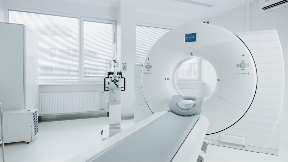
When entering the scanner, the person lies down, usually on their back, on a flat bed that goes into the donut-shaped PET scanner, sliding in and out of the scanner. The scan is painless and can take between 30 minutes to one hour.
MRI Scan
An MRI is a non-invasive, painless type of radiology imaging using radio waves and strong magnets that a powerful computer turns into detailed images of the inside of the body.
MRI scans can take between 15-90 minutes and can be done either with or without contrast. Most soft tissues can be seen well enough without contrast. However, in certain scenarios, contrast can help enhance the images.
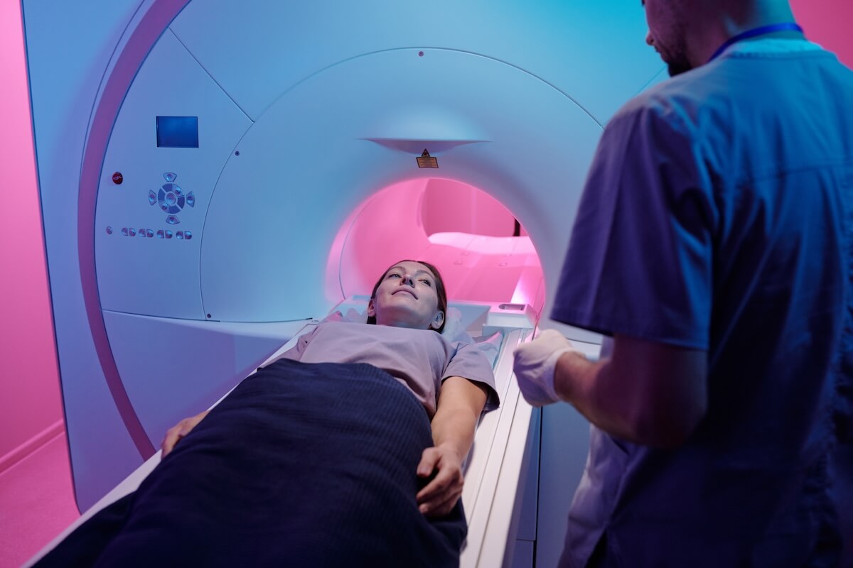
In a contrast MRI, the person is usually given a contrast dye called gadolinium, injected into their vein before the scan. To have the scan, the person lies down on their back on a flat bed, which is moved into the MRI scanner.
Once an MRI or PET scan has been carried out, the scans are reviewed by a radiologist or nuclear medicine physician who sends their findings to the requesting healthcare practitioner to be shared in a follow-up with the patient.
What Can a PET Scan vs. MRI Show?
A person may require a PET scan or an MRI based on their clinical need and which potential conditions are being looked for on the scan.
PET Scans
A PET scan can be a full body scan or imaging focused on one area of the body. It can help with the diagnosis of different conditions, such as:
- Different types of cancer
- Brain disorders, including dementia such as Alzheimer’s disease, brain tumors, and epilepsy
- Heart conditions like coronary artery disease (PET scans can pick up areas of decreased blood flow)
PET scans can also be useful in investigating a known cancer to see if it has spread or how well treatment is working. They can also be used to prepare for heart or brain surgery or to show where to take a biopsy.
Not all cancers will show up on a PET scan, and other imaging or tests might be needed to confirm if an area picked up by the tracer is cancerous or not. A PET scan can be carried out by itself or simultaneously as a CT scan (PET-CT scan) or an MRI (PET-MRI).
MRI Scans
MRI imaging can be used across most of the body and is particularly good at looking at soft tissues. An MRI can either be a full body scan or focus on a specific body area. Conditions MRI scans can look for include:
- Brain and spine: This includes multiple sclerosis, stroke, aneurysms, and brain and spinal cord tumors.
- Bone and joints: For example, primary bone tumors and joint or tendon injuries.
- Soft tissues: Visualizing organs, muscles, nerves, tendons and ligaments, including soft tissue sarcomas.
- Abdominal and pelvic organs: Including visualizing the liver, kidneys and pancreas and detecting tumors in the prostate, bladder, uterus, or ovaries.
- Heart and lungs: This includes structural and functional abnormalities and tumors and diseases affecting the heart muscle.
- Breasts: If clinically indicated, someone can have a dedicated breast MRI with a specialized breast coil, available at certain specialist units.
A specialized type of MRI called magnetic resonance angiography (MRA) can also measure blood flow through the various blood vessels in the body.
While these are diagnostic MRI scans, screening whole-body MRIs are available for those without symptoms (asymptomatic) who want to be proactive about their health.
What Is More Accurate: MRI or PET Scan?
It’s not a case of one type of scan being more accurate than another. A PET scan vs. MRI can be more useful in certain scenarios depending on the conditions being looked for. The two scans are also used together in a PET-MRI for certain situations, such as confirming whether an abnormality seen on a PET scan is likely to be cancerous (malignant).
PET Scan vs. MRI: Potential Risks and Challenges

Both scans are non-invasive (not including injection of the tracer or any MRI contrast), painless, and considered safe. There are, however, some potential risks, side effects, and challenges to know about.
Radiation
Only the PET scan gives exposure to radiation, and this is one of its biggest disadvantages. However, this radiation is in small amounts, and the risk of causing any issues such as cancer in the future is very small.
The radioactive tracer passes out of the body within a few hours and drinking lots of water can help pass it quicker. Those who have had a PET scan should avoid being around pregnant women and young children and babies for six hours after the scan.
The small risk from the radiation would be increased if a person has repeated PET scans, particularly if this was combined with CT scans, which also expose them to small amounts of radiation. However, the risks are still very small.
MRI scans do not give a person any radiation exposure. Therefore, this type of scan can be preferable for certain patients or annual scans.
Pregnancy
According to the American College of Obstetrics and Gynecologists, if someone is pregnant, MRI scans should be considered on a case-by-case basis after discussion with a healthcare practitioner. MRIs and ultrasounds are considered the safer options for pregnant women needing imaging, and MRIs without contrast are preferable.
PET scans would not be the first choice for a pregnant or breastfeeding woman due to radiation exposure. But again, if there is a clinical need, the decision would need to be discussed with a healthcare practitioner, weighing the risks and benefits.
Claustrophobia
If someone has claustrophobia, an MRI might be challenging as it occurs in a fairly small space. In some cases, a mild sedative can be requested ahead of the appointment day. An open MRI machine is also an option, if available, for those who struggle in confined spaces. However, open MRI images are lower resolution due to a weaker magnetic field, and 360 imaging may not be possible.
So which is more claustrophobic: a PET scan or MRI? A PET scan will likely be the easiest of the two to manage in this situation, as the scanner isn’t as long as the MRI scanner. Also, with a PET scan, the bed moves slowly in and out of the scanner rather than staying in the scanner the whole time.
Needing to Lie Still
For both scans, the person having the scan must remain still for much of the scanning time. This might be challenging for some people. If this is the case, the person should discuss it with the healthcare practitioner before the scan. Both scans last at least 15 minutes, so this may cause some discomfort even if the person is OK remaining still.
Metal
Due to the strong magnetic field used in an MRI, all metal items must be removed before the scan. The scan can’t be performed on patients with certain types of metal inside or on their body, including those with:
- Cochlear implants (for deafness)
- Pacemakers or implantable defibrillators (for heart rhythm abnormalities)
- Any metal fragments in the body (e.g., from an injury or dental bridge)
The person having the scan is given a detailed checklist and consent form to fill out before the scan so a decision can be made on whether the scan can proceed.
Allergic Reactions
It’s possible to have an allergy to contrast dye used in an MRI with contrast or the radiotracer used in PET scans, although this is rare. If someone has a known allergy, they should inform their healthcare practitioner before the scan.
Other side effects from the MRI contrast dye can include headache, dizziness, nausea and vomiting, or a skin rash. Rarely, someone can experience swelling and pain around the site where the PET radiotracer is put in if some leaks out.
Noise
Both scans can be noisy, therefore earplugs or headphones playing music are usually provided to protect hearing and provide a distraction. The person performing the scan can talk to the person being scanned and vice versa.
The Right Scan for the Right Situation
When choosing between a PET scan vs. an MRI scan, it all depends on the reason for having the imaging done. If you need a scan for diagnostic purposes, your healthcare practitioner can make the decision with you based on clinical need and talk you through the risks and benefits of the different options. If there’s an urgent need for a scan, any risks involved would likely be outweighed by the need to rule out the condition that is being investigated.
We have covered the pros and cons of the two different scans in terms of being used to aid diagnosis if someone has symptoms. However, if you’re considering having a proactive scan while you are symptom-free, an MRI is a great choice for a non-invasive scan without any risk of radiation exposure.
If you’d like to consider an Ezra Full Body MRI, you can easily book online. The scan takes as little as 30 minutes, and you’ll usually receive your results within 5-7 business days via an Ezra report that you may discuss with a healthcare professional via a telehealth visit.


