A magnetic resonance imaging (MRI) scan uses radio waves and magnetic fields to produce a highly detailed image of your body1. Your medical practitioner may order an MRI of the spine to try to diagnose whether there is an anatomical problem associated with persistent back pain.
This article will discuss when an MRI is important, what an MRI can find, and how to effectively prepare for an MRI.
Why Would a Doctor Order an MRI of the Spine?
The spine is crucial to movement, support, and everyday quality of life. Therefore, any issues with the spine can have a large impact on carrying out day-to-day activities. A doctor may order an MRI of the spine to identify the cause of any spinal issues that you may be experiencing, including2:
- Persistent or severe back pain
- Back pain with a fever
- Nerve compression symptoms (eg numbness, tingling, or weakness)
- Pain following injury to the back
- Stiffness in the back that restricts movement
A full-body MRI scan is a good way to obtain an overview of your spine, either to identify the root causes of any symptoms or as a pre-surgery process to help doctors prepare3.
What Does an MRI Scan of the Spine Show?
Normal Spine Anatomy
The spine is an S-shaped structure of bone starting at the base of your skull and ending at the tailbone. It comprises 33 vertebrae (small bones stacked together) connected by intervertebral disks and facet joints made of cartilage4. The vertebrae (except for the bottom two) can all move to support a range of motion. The disks and joints provide cushionning and support, enabling the vertebrae to slide against each other as you move. Protected within the spine is the spinal cord, a column of nerves extending from your brain down the full length of the spine. Nerves branch from the spinal cord between the vertebrae, carrying signals from the brain to the muscles4.
Abnormal Spinal MRI Results
MRIs form a detailed image of the spine and can help to differentiate between different common conditions that may present with similar symptoms. These conditions include tumors, herniated disks, hemorrhage, degenerative diseases (such as multiple sclerosis), spinal stenosis (narrowing of the spinal canal which can compress nerves), and infections3.
Limitations of MRI Findings
Although diagnostically helpful in many cases, MRIs also have limitations. False positives from MRI scans are relatively common, potentially showing the presence of abnormalities that are asymptomatic and unrelated to the current issue being investigated. MRIs cannot determine whether tissue appearing abnormal is functioning normally, or distinguish abnormalities causing pain from those causing no pain5. For example, MRI scans of a group of asymptomatic people found that 52 percent presented with at least one abnormality (eg a bulged disk)6. MRIs are best for imaging soft tissues, such as the spinal cord and discs, however, X-rays and CT scans are better for detailed imaging specifically of the bone3.
Does a Spinal MRI Show Organs?
A spinal MRI focuses on features of the spine, such as the vertebrae, discs, and the spinal cord. However, depending on the region of the spine being scanned, spinal MRIs can incidentally show organs. In particular, scans of the lumbar spine can provide images of organs, including the kidneys, liver, spleen, and uterus7. It is possible that this incidental imaging of organs may show abnormalities within these organs. These images may only capture part of an organ and may be obscured by image artifacts due to a spinal MRI focussing on the spine. In these situations, further imaging will be required to characterize any abnormalities in other organs7.

MRI in Different Regions of the Spine
What Can a Cervical Spine MRI Show?
The cervical spine is made up of 7 vertebrae and 8 nerve branches; it supports your head and allows movement, such as flexion, extension, and rotation, in addition to protecting the spinal cord and arteries carrying blood to the brain8. Cervical spine MRIs can identify abnormalities with the cervical spine, nerves, and adjacent soft tissues. These can include8:
- Cervical radiculopathy – cervical nerves being pinched by cervical vertebrae resulting in tingling, numbness, and pain
- Herniated discs – a tear or leak in a disc between vertebrae
- Bone spurs – growths on the cervical vertebrae
- Cervical degenerative disk disease – the wearing down of discs over time
- Cervical spondylosis – neck arthritis
What Can a Thoracic Spine MRI Show?
The thoracic spine is the longest section of the spine, found between the cervical and lumbar spinal regions. It is made up of 12 vertebrae and 12 nerve branches; it protects the spinal cord and branching nerves in addition to providing attachments for your ribs and stabilizing your rib cage9. The nerves branching from the thoracic spine transmit signals from the brain to major organs, including the lung, heart, and liver. MRI scans of the thoracic spine can identify abnormalities, including9:
- Spinal tumors – these can form anywhere along the spine (including in the spinal cord) but are most common in the thoracic and lumbar regions.
- Bulging discs and disc degeneration
- Cysts
- Thoracic radiculopathy – pinched nerves; this region of the spine is the least likely to experience this problem
- Infections and abscesses
- Compression fractures – the collapse of a vertebra causing severe pain; can result from trauma and conditions such as osteoporosis or a spinal tumor
What Can a Lumbar Spine MRI Show?
The lumbar spine is formed of 5 vertebrae and 5 nerve branches; it supports the majority of the body weight and provides stability and balance in addition to controlling movement in the lower limbs10. Lumbar spine MRIs can reveal anomalies, including10:
- Bulging or herniated discs
- Lumbar stenosis – narrowing of the spinal canal causing nerve compression; symptoms include pain or numbness in the lower body
- Spondylolisthesis – the slipping of a lumbar vertebrae out of place, putting pressure on a nerve
- Compression fractures – the collapse of a vertebra causing severe pain; can result from trauma and conditions such as osteoporosis or a spinal tumor
- Sciatica – nerve compression of the sciatic nerve
- Cauda equina syndrome – compression of a group of nerve roots called the cauda equina; symptoms include incontinence
- Spinal tumors
- Unusual spine curvature – including lumbar lordosis (excessive curvature of the spine) and scoliosis
Does a Lumbar Spine MRI Show the Pelvis?
Due to the proximity of the lumbar spine to other areas of the pelvis, parts of the pelvis will likely be imaged as part of a lumbar spine MRI scan. However, the pelvis is not the main focus of these scans, so only certain areas of the pelvis may be captured. In addition, image artifacts may obscure regions of the pelvis, as the radiographer will be focussing on obtaining an optimized image of the lumbar spine, not the pelvis. Incidental findings in the pelvic regions are possible. For example, in one study, gynecological findings (such as ovarian cysts and endometrioma) were discovered in 14 percent of patients undergoing a lumbar MRI11. However, if a detailed image of the pelvis is needed, an MRI focusing on the pelvis should be carried out.
Preparing for an MRI Scan of the Spine
A spinal MRI requires very little preparation. Unless told otherwise, you will be able to eat, drink, and take medications as usual on the day of your MRI. In certain circumstances, you may be required to stop eating and drinking for up to 4 hours12 before the scan – medical professionals will inform you whether this is required.
Prior to the scan, a thorough screening will be carried out to ensure that you do not have any metal within your body that could interfere with or cause damage to the scan (the majority of metal within your body, for example, joint replacements, will be MRI-compatible)13. Any external metal objects, such as watches, jewelry (including piercings), dentures, and hearing aids, must be removed. It is also important to inform your healthcare professionals of tattoos as these may contain metal which can heat up in the MRI scanner, causing burns14. The wearing of make-up should be avoided as this can occasionally contain magnetic substances which can result in poor quality MRI images15.
You should inform your healthcare provider of any allergies and known health conditions, especially if these involve your kidneys. This is particularly important if an MRI with contrast is being considered. MRIs with contrast may cause mild side effects and could result in an allergic reaction to the contrast dye16.
If you have claustrophobia, talk with your medical care team.H3: What Should I Wear for an MRI Scan of the Back? When attending an MRI scan, you should wear comfortable and loose clothing and shoes that are easy to remove. You may be given a gown or pajamas to change into at the imaging facility. Clothes containing metal, such as zippers and underwired bras, should be avoided as the metal may interfere with the imaging equipment and decrease the quality of the MRI image17.
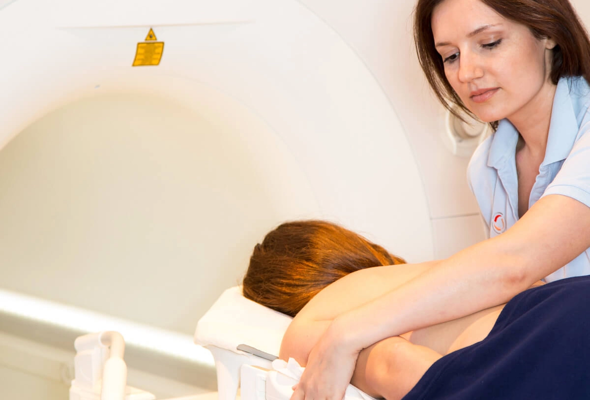
MRI of the Spine: What To Expect From the Scan
An MRI scan is generally an outpatient procedure. You will lie on a table which will slide into the scanner. When the scanning is in process, the MRI machine will make loud thumping and clicking noises; this is completely normal13. You will often be given a pair of earplugs to protect your ears, some of our Ezra imaging locations will provide headphones with music playing throughout the scanning experience. Once the scan is complete, you are free to leave and carry out the rest of your day as usual. An expert radiologist will analyze the images obtained from the MRI and will produce a report that will be communicated to the patient by a specialist, who will also be able to discuss any next steps based on the results.
How Long Does a Full Spine MRI Take?
An MRI scan generally takes between 15 and 90 minutes, depending on the size of the area being scanned. The total time in the scanning facility (though not the scanner itself) will be increased if a contrast agent is required. Ezra scans take only 1 hour for their full-body scan.
Does Your Whole Body Go In for a Lower Back MRI?
Full-body MRI scans will require your whole body to be within the scanner. However, lower back (lumbar) spine MRIs often do not. In these situations, you will be put into the scanner feet-first and only the lower part of your body will have to enter the machine, leaving your head and upper body outside of the scanner. This can be more comfortable for those who experience claustrophobia in MRI scanners18.
Is an MRI Better Than Other Scans?

The form of imaging technique used in medical scans is dependent on what is being scanned. When choosing the type of scan to be performed, medical professionals will assess the risk-benefit ratio. MRI scans, CT scans, and X-rays are all painless and non-invasive – enabling doctors to see inside your body without having to cut it open19. MRI scan images are often better quality, in particular when imaging soft tissue, however, CT scans are preferred in certain areas of the body where unavoidable movement (eg movement of the chest from breathing) may cause issues with MRI images. CT scans involve greater risks, as the patient is exposed to ionizing radiation. While the amount of radiation dose you get from a single CT scan is small, regular screening with CT scans can increase the risk of developing cancer20. MRIs do not involve radiation and are therefore considered safer13.
Choosing an MRI Center
Choosing a high-quality scanning facility is essential to obtaining helpful information from an MRI scan. Facilities accredited by reputable organizations, such as the American College of Radiology21, will follow stringent quality and safety standards, ensuring that you receive the best possible experience. In addition, choosing facilities that use advanced technologies and have experienced radiologists will ensure that you receive high-quality images and expert scan interpretations. Ezra always partners with top imaging centers and radiologists.
Summary: What Does an MRI Scan of the Spine Show?
Maintaining spinal health is key to long-term quality of life. An MRI of the spine is a valuable tool to assist medical professionals in diagnosing a wide range of conditions that may affect the spine, including spinal tumors, herniated discs, and nerve compression. MRI scans are painless and non-invasive, requiring minimal preparation and time to complete.
By getting an annual Ezra full-body MRI scan as part of your routine health care, you can monitor changes in your health over time and detect possible abnormalities like early cancer or other diseases earlier.
When a problem is detected in the early stages, your treatment can be more effective.
The Ezra full-body MRI scans up to 13 organs for signs of potential cancer. It is priced at $2395. Our more advanced service, the Ezra Full Body Plus, scans 14 organs – including the lungs – and is priced at $2695.
Understand your risk for cancer with our 5 minute quiz.
Our scan is designed to detect potential cancer early.
References
1. Magnetic Resonance Imaging (MRI). May 22, 2024. Accessed February 6, 2025. https://www.hopkinsmedicine.org/health/treatment-tests-and-therapies/magnetic-resonance-imaging-mri
2. British Association of Spine Surgeons - The Spine and MRI Scanning. Accessed February 6, 2025. https://spinesurgeons.ac.uk/The-Spine-and-MRI-Scanning
3. Magnetic Resonance Imaging (MRI) of the Spine and Brain. May 28, 2024. Accessed February 6, 2025. https://www.hopkinsmedicine.org/health/treatment-tests-and-therapies/magnetic-resonance-imaging-mri-of-the-spine-and-brain
4. Function of the Spine. Cleveland Clinic. Accessed February 6, 2025. https://my.clevelandclinic.org/health/body/10040-spine-structure-and-function
5. MRI: Understanding its limitations | British Columbia Medical Journal. Accessed February 6, 2025. https://bcmj.org/articles/mri-understanding-its-limitations
6. Jensen MC, Brant-Zawadzki MN, Obuchowski N, Modic MT, Malkasian D, Ross JS. Magnetic resonance imaging of the lumbar spine in people without back pain. N Engl J Med. 1994;331(2):69-73. doi:10.1056/NEJM199407143310201
7. Incidental Findings on Musculoskeletal MR | Radsource. October 3, 2005. Accessed February 6, 2025. https://radsource.us/incidental-findings-on-musculoskeletal-mr/
8. Cervical Spine (Neck): What It Is, Anatomy & Disorders. Cleveland Clinic. Accessed February 6, 2025. https://my.clevelandclinic.org/health/articles/22278-cervical-spine
9. Thoracic Spine: What It Is, Function & Anatomy. Cleveland Clinic. Accessed February 6, 2025. https://my.clevelandclinic.org/health/body/22460-thoracic-spine
10. Lumbar Spine: What It Is, Anatomy & Disorders. Cleveland Clinic. Accessed February 6, 2025. https://my.clevelandclinic.org/health/articles/22396-lumbar-spine
11. Ebru Hasbay MS. Incidental Gynecological Findings in Lumbar Spine MRI: The Prevalence of These Findings and Their Importance For Patient Management. doi:10.4274/terh.galenos.2023.01643
12. MRI scan - How it’s performed. nhs.uk. September 5, 2018. Accessed February 6, 2025. https://www.nhs.uk/conditions/mri-scan/what-happens/
13. Health C for D and R. Benefits and Risks. FDA. Published online September 2, 2019. Accessed February 6, 2025. https://www.fda.gov/radiation-emitting-products/mri-magnetic-resonance-imaging/benefits-and-risks
14. Ross JR, Matava MJ. Tattoo-Induced Skin “Burn” During Magnetic Resonance Imaging in a Professional Football Player. Sports Health. 2011;3(5):431-434. doi:10.1177/1941738111411698
15. Smith FW, Crosher GA. Mascara--an unsuspected cause of magnetic resonance imaging artifact. Magn Reson Imaging. 1985;3(3):287-289. doi:10.1016/0730-725x(85)90359-5
16. Gadolinium Side Effects and Toxicity Explained. Accessed February 6, 2025. https://www.drugwatch.com/gadolinium/side-effects/
17. MRI scan - How it’s performed. nhs.uk. September 5, 2018. Accessed February 6, 2025. https://www.nhs.uk/conditions/mri-scan/what-happens/
18. Claustrophobia and MRI. UCSF Radiology. May 29, 2013. Accessed February 6, 2025. https://radiology.ucsf.edu/patient-care/prepare/claustrophobia-mri
19. CT Scan Versus MRI Versus X-Ray: What Type of Imaging Do I Need? July 9, 2024. Accessed February 6, 2025. https://www.hopkinsmedicine.org/health/treatment-tests-and-therapies/ct-vs-mri-vs-xray
20. Mathews JD, Forsythe AV, Brady Z, et al. Cancer risk in 680 000 people exposed to computed tomography scans in childhood or adolescence: data linkage study of 11 million Australians. BMJ. 2013;346:f2360. doi:10.1136/bmj.f2360
21. American College of Radiology. Accessed February 6, 2025. https://www.acr.org/


