
Key takeaways:
- Advances in medical imaging provide for better and faster prostate cancer detection. A prostate MRI is more accurate in finding prostate cancer, than digital rectal exams or PSA tests, which are inconclusive.
- The American Cancer Society estimates 191,930 new cases of prostate cancer in 2020. The five-year survival rate is 98%.
- Prostate cancer may not cause symptoms for years, even with elevated PSA levels.
- PSA testing can help flag the need for further investigation, but it also can lead to overdiagnosis or overtreatment.
Increasingly, doctors are turning to prostate MRI scans to detect prostate cancer. That’s because MRI technology improvements are leading to more accurate detection.
A correct screening test such as a prostate MRI can help patients minimize overdiagnosis, have a more targeted MRI-guided biopsy, or monitor the progression of disease.
Doctors may also use this advanced technology to help diagnose infections such as prostatitis or an enlarged prostate (sometimes referred to as benign prostatic hyperplasia (BPH), which is relatively common in men over 50 and usually does not represent a severe medical condition.
If prostate cancer is detected, doctors can use prostate MRI technology to look at the extent of prostate cancer and ascertain whether it has spread to other parts of the body by metastasis.
About Prostate Cancer
According to the American Cancer Society’s estimates, 191,930 American men will have contracted prostate cancer in 2020. After skin cancer, the most common cancer in men in the United States is prostate cancer. And while a diagnosis of prostate cancer is distressing, there’s reason for hope.
Prostate cancer grows slowly and may not ever cause noticeable symptoms, or may not cause them for years.
During a routine digital rectal examination (DRE), abnormalities often lead to a prostate-specific antigen (PSA) blood test.
PSA Testing May Not Be an Accurate Indicator of Prostate Cancer
PSA testing remains controversial, as it can lead to overdiagnosis and overtreatment.
A PSA level of >4 ng/mL isn’t necessarily indicative of prostate cancer, which should be confirmed by a prostate MRI.
If you have an elevated PSA level (>4 ng/mL), your doctor will likely recommend an MRI scan to get a more accurate diagnosis because of these PSA testing drawbacks:
- Medications for benign prostatic hyperplasia (BHP) or an enlarged prostate gland can skew your PSA levels.
- There’s a moderate risk of false-positive or false-negative test results with PSA. Approximately 15% of men with a PSA of <4 ng/mL will be diagnosed with prostate cancer following a biopsy. Even with an elevated PSA of >4 ng/mL, 75% of men will not have cancer, and may undergo an unnecessary prostate biopsy.
- Reliance on PSA levels alone can lead to overdiagnosis and overtreatment.
Prostate MRI Scans Can More Accurately Detect Prostate Cancer
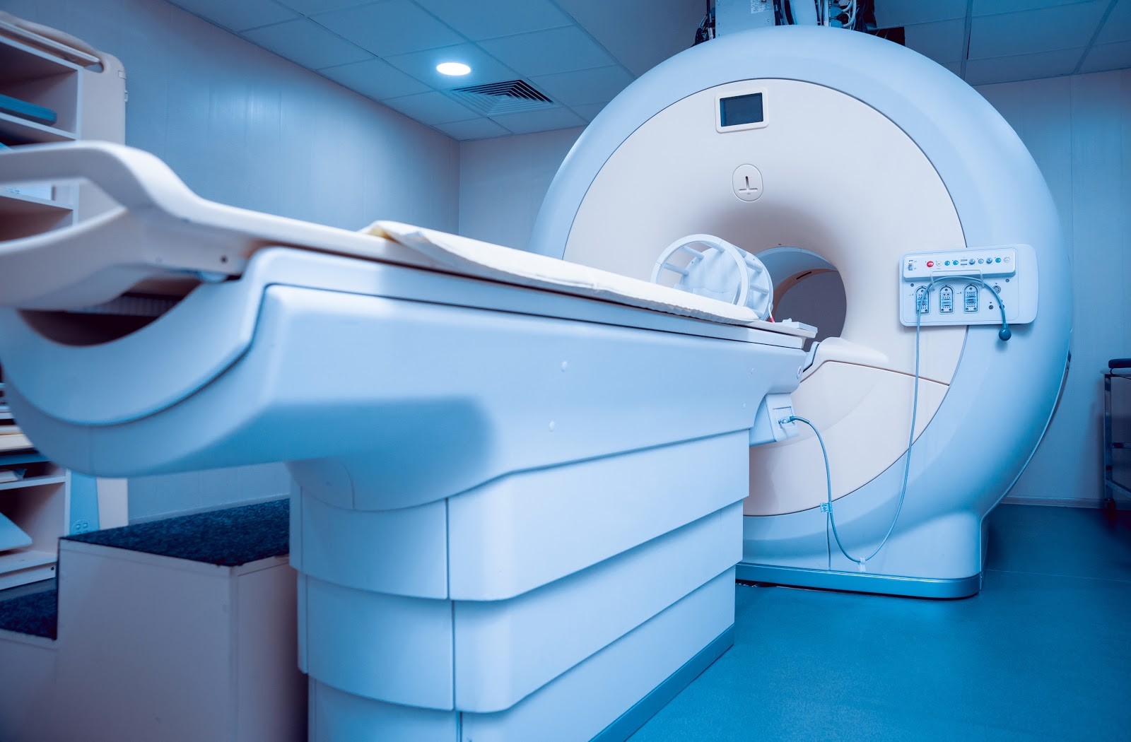
Before undergoing a prostate biopsy, your doctor may want you to have a prostate MRI scan to confirm a tumor’s presence. Magnetic resonance imaging (MRI) is safe, non-invasive, and pain-free.
Screening with the latest prostate MRI technology can detect more than 90% of clinically-significant prostate cancers.
An MRI uses radio waves in a strong magnetic field to create detailed images of the body that differentiate between normal and cancerous tissues.
A full-body MRI usually takes 60 minutes. After scanning the body with the MRI machine, the radiologist reviews the images and writes a report for your doctor. A prostate MRI done with ezra takes less than 20 minutes.
An MRI provides better soft-tissue contrast to highlight the differences between fat, water, muscle, and other soft tissue, as well as water molecule motion (water diffusion) and blood flow (perfusion imaging).
With a contrast agent such as gadolinium-based contrast agents (GBCAs), the radiologist can see any abnormal prostate tissue in more detail. (Note: ezra does not use contrast for our screening prostate MRI)
A radiologist uses PI-RADS (Prostate Imaging–Reporting and Data System) to evaluate and report suspected prostate cancer.
If the radiologist thinks it’s necessary, they will recommend further investigation and, perhaps, a prostate biopsy to obtain a cancer diagnosis before deciding on the best next steps for the patient..
Advances in MRI Technology
A prostate MRI scan is often the first step in following up on a suspected prostate cancer diagnosis.
The strength of the MRI machine’s magnetic field is measured in “teslas.”
A tesla is a measurement unit that is used to define the magnetic flux density. With higher tesla scanners, the magnetic field is stronger, which results in higher quality images
The magnet and its magnetic field is the essential aspect of an MRI scanner.
Standard MRI scanners operate at 1.5 Tesla. Many radiology facilities now use stronger machines, going up to a 3.0 Tesla MRI.
A 3.0 Tesla MRI provides a bigger signal-to-noise proportion, which is a significant factor in producing the best quality picture. The strength of a 3 Tesla MRI has numerous benefits for radiologists and their patients.
Nowadays, an MRI scan can produce 3-D images, much like a CT scan.
Magnetic resonance spectroscopic imaging (MRSI) is increasingly used, as it is a high-quality evaluation tool for determining the extent and aggressiveness of primary and recurrent prostate cancer.
A non-invasive procedure, MRSI utilizes an endorectal coil. An MRSI measures biochemical changes in the prostate where tumors are present. An MRSI offers more data about cellular activity and involvement and better detects tumor volume (size). (Note: ezra uses a flat-sheet abdominal coil instead of an endorectal coil, which improves the patient experience).
The information gained from an MRSI helps with staging prostate cancer.
Multiparametric-magnetic resonance imaging (mpMRI) helps radiologists quantify and standardize tumor images. The advantage is that mpMRI leads to improved image interpretation accuracy and reporting of the scan results, based on its three-dimensional nature.
For oncologists and their patients, mpMRI (or Mp-MRI) images are more accurate in diagnosing, locating, classifying, and staging clinically significant prostate cancer, according to one article in PubMed Central (PMC).
An mpMRI can be helpful in seeing the difference between low-risk or slow-growing prostate cancer and high-risk (aggressive) prostate cancers.
An mpMRI has become the go-to modality for detecting areas thought to be prostate cancer. It also provides a great assist to targeted biopsies.
There are three methods for performing a prostate biopsy:
- In-bore MRI prostate biopsy is performed while the patient is in the MRI machine.
- Cognitive fusion biopsies use MRI images to estimate the location of a lesion or prostate gland tumor during ultrasound procedures.
- Software-based fusion displays MRI and TRUS images together to guide doctors with targeted biopsies.
Magnetic Resonance Imaging (MRI) and Other Diagnostic Tools for Better Prostate Screening

As part of reaching a more conclusive diagnosis, your doctor may order a transrectal ultrasound (TRUS), also known as a prostate sonogram or endorectal ultrasound.
The procedure involves a rectal probe that bounces high-energy sound waves off internal organs and tissue to produce a sonogram.
TRUS looks for abnormalities in the rectum and nearby structures, including the prostate. During the biopsy, a urologist usually collects 12 core samples from suspicious areas of the prostate, guided by a fusion of MRI and TRUS images.
Ultrasound-MRI fusion-guided needle biopsies have shown improvements in targeting and sampling suspicious regions of the prostate.
Prostate Cancer Is Rarely Terminal, Especially When Detected Early
The second most common cancer, a prostate cancer diagnosis is not hopeless. Prostate cancer is usually a slow-growing cancer, and those diagnosed with it can have a good quality of life. They may not ever show symptoms.
Prostate MRI screening helps doctors and their patients make more informed decisions about treatment choices.
Advances in MRI technology are helping doctors segment the prostate gland and lesions and become more efficient at locating tumors and lesions in the prostate.
Get your ezra Full Body MRI Scan — it only takes one hour and it screens for potential early cancer and early disease in up to 13 organs, including the prostate.
Sign up for a consultation today.


