Breast imaging techniques are essential for monitoring breast health, especially for detecting cancer within breast tissue. Breast cancers have a far better prognosis when caught early, which makes engaging with screening programs crucial for maintaining long-term health. Fortunately, several powerful technologies are available for breast imaging, and screening programs have been shown to improve survival rates. It’s completely normal to be anxious about going for breast screening; however, knowing what to expect when you go for different types of screening can help alleviate some of these concerns.
The article will focus on understanding breast magnetic resonance imaging (MRI) and ultrasound. It will cover how these techniques compare to each other and how this information can help you and your healthcare provider select the best imaging option for you should the need arise.
What to Expect from Breast MRI and Ultrasound Procedures: A Detailed Overview
What is a Breast MRI?
Mammographies are commonly used for breast cancer screening, but MRIs are also used in some circumstances. Let’s look at how MRI scans work and how they are used for breast cancer screening.
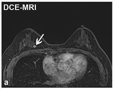
How Breast MRI Works
MRI is a medical imaging technique that uses strong magnets and radio waves to create detailed images of the inside of the body without using radiation. MRIs can pick up on minor abnormalities in breast tissue that mammographies can sometimes miss. MRI technology can be particularly useful for examining dense breast tissue, which is associated with a higher risk of cancer and makes finding tumors more difficult with mammography.
When is a Breast MRI Recommended?
Breast MRIs are often recommended for screening where there is a high risk of cancer or when dense breast tissue makes mammography less viable. MRIs are also employed when cancer has already been diagnosed to monitor disease progression and treatment efficacy. When results from other imaging techniques like mammography or ultrasounds are inconclusive, an MRI can help by providing more detailed images. MRIs are also used to assess the integrity of breast implants.
What to Expect During a Breast MRI
Before the scanning begins, you will be asked to remove any metal objects, remove your clothes, and put on a gown. During the scan, you will be asked to lie face down in the MRI machine, and the scan will typically take 30 to 60 minutes. An MRI is a non-invasive procedure that does not involve compression of the breasts. However, contrast agents are often used to improve image quality, and they are administered via injection. It’s important to discuss any concerns you may have about the procedure, such as claustrophobia or other discomfort, with your healthcare provider.
What is a Breast Ultrasound?
Like MRIs, ultrasounds are usually secondary to mammography for routine screening, though they are often used together.
How Breast Ultrasound Works
Ultrasound is an imaging technique that uses sound waves to take detailed images of the body, including breast tissue. Ultrasound equipment sends sound waves into the breast, and as they bounce back, they create images of the breast tissue, allowing doctors to see inside without using ionizing radiation. Ultrasound effectively evaluates lumps or other areas of concern that may be found during physical examination but do not always appear on mammograms.
When is a Breast Ultrasound Recommended?
Breast ultrasound is useful in many circumstances, such as evaluating suspicious areas detected by other imaging techniques, helping to guide biopsy needles, and imaging dense breast tissue that is hard to analyze with mammography. Ultrasounds also help to provide additional information about breast tissue, such as distinguishing between solid masses and fluid-filled cysts.
What to Expect During a Breast Ultrasound
During a breast ultrasound, you will be asked to lie on an examination table, and a special gel will be applied to the breast area. The procedure is carried out by a sonographer using a handheld instrument called a transducer, which emits the sound waves that create the images. Ultrasounds are painless, noninvasive procedures that typically take 15 to 30 minutes to complete.
Comparing Breast MRI and Ultrasound
Knowing the differences between breast MRI and ultrasound is important for choosing the best screening option and for understanding how they complement each other and mammography.
Differences in Imaging Techniques
MRIs provide more detailed images of breast tissue than ultrasounds. However, ultrasounds are effective in examining lumps and other problem areas. While both procedures are non-invasive and do not involve exposure to ionizing radiation, MRIs can cause discomfort in some patients.
Complementary Roles in Breast Imaging
Together with mammography, breast MRI and ultrasound provide a comprehensive suite of techniques for examining breast tissue. When used in conjunction, both can cover the limitations of mammography and provide healthcare professionals with more information. These techniques allow screening procedures to be tailored to patients' needs. For instance, the American Cancer Society recommends MRI for screening high-risk individuals, while ultrasounds are more common in regular diagnostic assessments alongside mammography.
Choosing the Right Procedure
The best choice of screening procedure depends on many parameters, including patient risk factors, age, breast density, and the specific clinical question healthcare professionals are trying to address. For example, high-risk individuals such as those with BRCA1 or BRCA2 mutation can benefit from MRI screening, which may pick up smaller, early-stage tumors. If a patient discovers a lump during self-examination, ultrasound can offer a safe, non-invasive way to investigate it. Be sure to discuss the available screening options with your healthcare provider.
The Benefits of Breast MRI and Ultrasound
Early Detection and Diagnosis
The earlier breast cancer is detected and diagnosed, the better the prognosis. Engagement with screening programs has been shown to significantly reduce breast cancer-associated mortality. All breast imaging technologies, including MRI and ultrasound, play an important role in ensuring that breast cancer is caught as early as possible.
Non-Invasive and Safe Procedures
It’s important to emphasize that both breast MRI and ultrasound are non-invasive and safe procedures with minimal associated risks. Unlike CT scans and X-rays, these techniques do not use harmful ionizing radiation. This makes them particularly useful for repeated screening, which benefits high-risk individuals for whom repeated screening is recommended. While these techniques are safe, going for a scan is a source of anxiety for most people. Discussing the scanning procedures and relaxation techniques with your healthcare provider can help alleviate your concerns.
Personalized Screening and Monitoring
Medical care is becoming increasingly personalized, which means that screening procedures can be tailored to an individual's specific needs rather than a one-size-fits-all approach. Personalization in healthcare requires flexibility and access to a wider range of technologies, enabling providers to better assess their patients. MRIs and ultrasounds provide this flexibility, allowing clinicians to address patient-specific questions and gain more insights into breast health than with one method alone.

Conclusion
MRI and ultrasound are important tools that, together with mammography, allow for detailed imaging of breast tissue. Each method has its strengths, with MRIs excelling in providing highly detailed images, and ultrasounds providing insights into lumps and other abnormalities. Both can be used to image dense breast tissue, which is challenging with mammography techniques. Discussing screening options with your healthcare provider, including any concerns you have, can help direct appropriate care and allow early breast cancer detection.
Advances in breast cancer imaging technology mean it’s becoming easier for clinicians to detect tumors. However, it’s important to remember that these advances are only beneficial when individuals engage with screening programs to help secure their long-term breast health.
If you want to be proactive about your health, why not book an Ezra MRI Scan? Our annual scan catches potential cancer earlier, leveraging AI through the screening process to make it more efficient, affordable and faster.


