Diagnostic imaging tests are fundamental to modern healthcare, offering non-invasive methods to visualize the body’s internal structures. These tests, including X-rays, CT scans, MRI, and ultrasound, use various technologies to create detailed images that aid in diagnosing, monitoring, and treating various medical conditions.
These techniques are crucial in early disease detection, accurate diagnosis, and effective treatment planning. From identifying fractures to detecting tumors, they are indispensable tools in medical practice.
This article aims to provide a comprehensive overview of diagnostic imaging tests, exploring their functions, applications, safety considerations, and recent technological advancements.
What is A Diagnostic Imaging Test? A Complete Overview
What Are Diagnostic Imaging Tests?
Diagnostic imaging tests are medical procedures that allow healthcare providers to visualize the inside of a patient’s body to identify, diagnose, and monitor various medical conditions. Using different technologies and techniques, these tests create detailed images of internal structures, organs, and tissues. The primary purpose of diagnostic imaging is to provide doctors with crucial information about a patient’s health without requiring invasive exploratory procedures.
These tests work by capturing images of the body’s interior using various forms of radiation, such as X-ray and computed tomography (ionizing), magnetic resonance, and ultrasound (non-ionizing). By producing clear and detailed images of specific areas within the body, diagnostic imaging tests enable healthcare professionals to detect abnormalities, assess the severity of conditions, and monitor treatment progress.
Diagnostic imaging is vital in modern medicine. It allows for earlier and more accurate diagnoses and improved treatment planning. These tests are essential for preventive care, disease management, and medical interventions across various specialties.
Types of Diagnostic Imaging Tests
X-rays
X-rays are one of the oldest and most widely used forms of diagnostic imaging. They work by passing low-dose radiation through the body, which is absorbed differently by various tissues based on their density. Denser tissues, like bones, appear white on X-ray images, while softer tissues appear in shades of gray.
X-rays are commonly used to:
- Detect bone fractures and joint dislocations
- Identify dental problems, such as cavities
- Diagnose chest conditions like pneumonia
- Locate foreign objects in the body
- Guide orthopedic surgery procedures
- Diagnose digestive problems
X-rays are quick, painless, and relatively inexpensive, making them valuable tools in emergency and routine medical care. However, they do involve exposure to small amounts of ionizing radiation, so healthcare providers carefully consider their use.
Computed Tomography (CT) Scans
Computed tomography (CT) scans use specialized X-ray equipment and computer processing to create detailed cross-sectional images of the body. The scanner rotates around the patient, taking multiple X-ray images from different angles, which are combined to produce 3D representations of internal structures.
Applications of CT scans include:
- Diagnosing and staging cancers
- Detecting internal injuries and bleeding
- Identifying bone and muscle disorders
- Guiding medical procedures like biopsies
- Planning radiation therapy or surgery
Advantages of CT scans include their ability to visualize soft tissues, blood vessels, and organs with exceptional clarity. However, CT scans expose patients to higher levels of radiation compared to conventional X-rays, which may slightly increase the lifetime risk of developing cancer. Despite this risk, when used appropriately, the benefits of CT scans generally outweigh the potential risks.
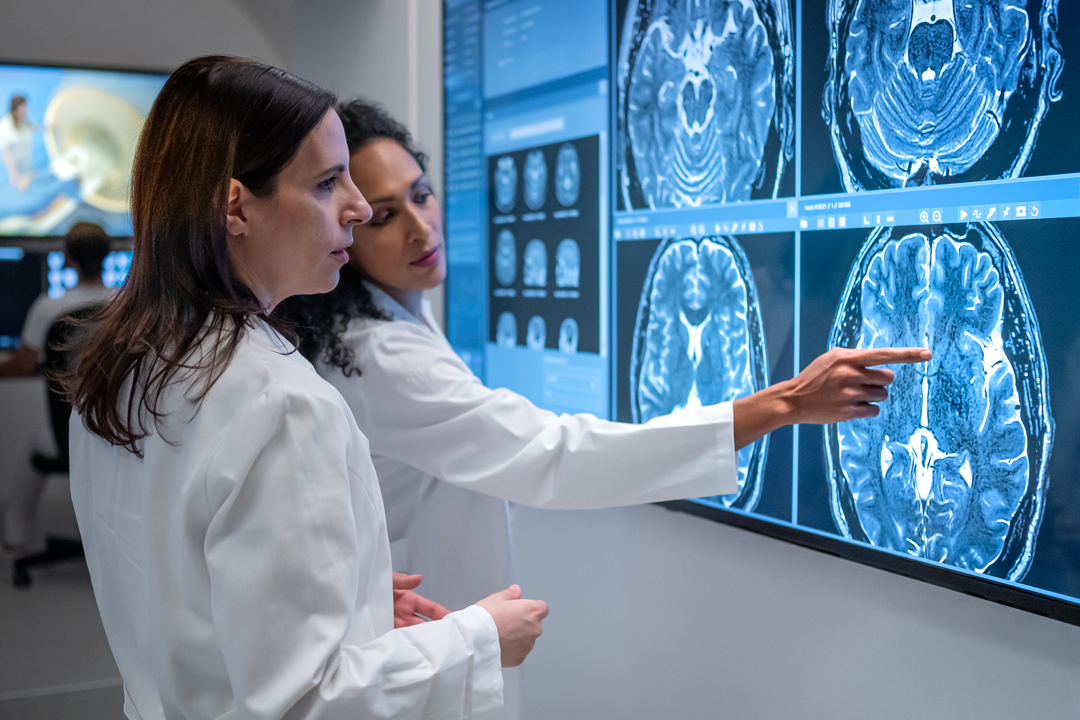
Magnetic Resonance Imaging (MRI)
Magnetic resonance imaging (MRI) scans use strong magnetic fields and radio waves to produce detailed images of the body’s internal structures. The technology works by aligning protons in the body with a magnetic field and then using radio waves to disturb this alignment. When the radio waves are turned off, the protons realign, emitting signals captured and converted into images by a computer.
MRI is beneficial for diagnosing soft tissue injuries, such as ligament tears and muscle damage, as well as brain conditions, including aneurysms, brain injury, tumors, multiple sclerosis, and strokes.
MRI scans are safer for repeated use because they are noninvasive and lack ionizing radiation. Furthermore, they provide better soft tissue contrast than other imaging techniques and can more easily show the difference between fat, water, and muscle. However, considerations include high cost, longer scan times, and potential issues for patients with metal implants or claustrophobia.
Ultrasound
Ultrasound imaging uses high-frequency sound waves to create real-time images of internal body structures. A transducer emits sound waves that bounce off tissues and organs, creating echos that are converted into visual images.
Uses include:
- Monitoring fetal development during pregnancy
- Examining organs like the heart, liver, and kidneys
- Guiding needles in biopsies or tumor treatments
- Assessing blood flow in vessels
- Examining lumps in the breast
- Evaluating levels of joint inflammation
Advantages include its safety (no ionizing radiation), real-time imaging capability, and portability. It’s also relatively inexpensive and widely accessible. However, it is less effective in imaging air-filled organs or structures behind bone, and the quality of images can be affected by patient body composition and technician skill.
How Diagnostic Imaging Tests Aid in Diagnosis
MRI and CT scans play important roles in cancer screening and diagnosis. MRI excels at producing detailed images of soft tissues, making it particularly effective for detecting tumors in the brain, spine, and other organs. It can accurately determine tumor size, location, and whether cancer has spread.
CT scans are invaluable for identifying cancers in the chest, abdomen, and pelvis. They offer quick and comprehensive imaging results, and they are especially useful for detecting lung nodules and abdominal masses.
Both techniques are essential for early cancer detection, accurate staging, and treatment planning. They allow healthcare providers to visualize internal structures non-invasively, guiding decisions on biopsies, surgeries, and other interventions. At Ezra, we provide a Full Body Plus scanning service that uses MRI and CT to comprehensively view your overall health. Ezra is for screening purposes only and should not be used as a diagnostic test.
Safety and Risks of Diagnostic Imaging Tests
Diagnostic imaging tests are generally safe when performed by trained professionals. However, some tests carry potential risks that healthcare providers carefully manage.
X-rays and CT scans involve exposure to ionizing radiation, which, in high doses, can increase cancer risk. Providers use the lowest effective radiation dose to mitigate this and limit unnecessary scans. MRI scans don’t use radiation but employ strong magnetic fields, requiring careful screening for metal implants or devices. Contrast agents used in some imaging procedures may cause allergic reactions in rare cases.
Overall, the benefits of diagnostic imaging typically outweigh the risks involved.
Advancements in Diagnostic Imaging
Recent advancements in diagnostic imaging technology are revolutionizing the field, significantly improving diagnostic accuracy and patient outcomes. One significant development is the integration of artificial intelligence (AI) into imaging. AI algorithms, particularly deep learning, enhance the interpretation of medical images by identifying subtle abnormalities that may be missed by human eyes, thus improving early disease detection and diagnostic precision. AI also aids in image segmentation and analysis, which is important for treatment planning and monitoring.
Another significant advancement is 3D imaging, which provides detailed three-dimensional views of internal structures. This enhances the visualization of complex anatomical areas and improves surgical planning and outcomes. These technologies increase the accuracy of diagnoses and streamline workflows, reduce human error, and offer personalized treatment options, ultimately leading to better patient care.

Summary: What Is A Diagnostic Test?
Diagnostic imaging tests have revolutionized modern healthcare, offering non-invasive methods to visualize the body’s internal structures. These technologies, from X-rays and CT scans to MRI and ultrasound, provide healthcare providers with powerful tools for early disease detection, accurate diagnosis, and effective treatment planning. Each imaging technique has unique strengths for addressing various medical conditions. While these tests generally carry minimal risks, healthcare providers carefully weigh the benefits against potential concerns such as radiation exposure in X-rays and CT scans. As medical imaging plays an increasingly important role in healthcare, the potential for individuals to be proactive about their healthcare becomes easier.
If you want to be proactive about your health, why not book an Ezra Full Body Plus scan? It utilizes MRI and CT to catch potential cancer earlier, leveraging artificial intelligence through the screening process to make it more efficient, affordable, and faster. Ezra is for screening purposes only and should not be used as a diagnostic test.


