
Regular health screenings are a vital component of proactive medicine, offering early detection of potential issues before symptoms appear1. Among these, multi-organ scans have emerged as a comprehensive tool for assessing overall health. Utilising advanced imaging technologies such as computed tomography (CT) and magnetic resonance imaging (MRI), multi-organ scans can identify early signs of serious conditions, including cancer.
In this article, we will discuss what multi-organ scans entail, what they can show, and their benefits. We’ll also help you make an informed decision about whether to start annual screenings to assess your overall health, so you can act proactively to live longer and better.
What Are Multi-Organ Scans?
Multi-organ scans are medical imaging procedures designed to reveal what’s happening inside your body before any symptoms appear. They can help clinicians detect signs of serious diseases, such as cancer, at early stages when they are more easily treatable.
At Ezra, we use MRI and CT technologies to provide detailed images of organs, from the brain to the pelvis. While both techniques can visualise the inside of your body, CT scans are more effective for some organs, and MRI is better suited for others.
CT Scan
CT is an advanced imaging method that utilises X-rays to generate detailed cross-sectional images of the body2. This technique has been widely used for many years, with advances in technology and research continually enhancing the safety, sensitivity, and accuracy of CT scans, thereby improving patient outcomes3.
During a CT scan, an X-ray tube rotates around the patient, emitting a focused X-ray beam that traverses the body. The scanner measures X-ray attenuation by detecting the amount of X-rays passing through tissues. This data is used to create images based on the varying absorption rates of different tissues. By capturing images from multiple angles, the scanner produces comprehensive cross-sectional views of the body, depicting tissue density on a scale known as the Hounsfield scale. Organs exhibit various densities; for example, a bone has a denser structure than soft tissue due to its higher calcium content. As a result, the different tissues will appear with varying contrasts on the scan4.

Your radiologist can then use your CT scan results to assess your organ’s health and identify abnormalities in specific organs and tissues. CT scans are especially beneficial for lung, heart, and abdominal screening.
- In your lung, a low-dose CT (LDCT) scan is the gold standard imaging procedure used in screening programs to detect early-stage lung cancer. Early detection of cancerous nodules can save your life; a national targeted lung cancer screening trial showed CT screening reduced lung cancer mortality by 26 percent in men and between 39 percent and 61 percent in women5.
- In your heart, a CT scan can detect aortic aneurysms and calcium within plaques in the coronary arteries6.
- A CT scan of the abdominal region can show kidney stones, cysts, enlarged lymph nodes, large masses, or an enlarged spleen and assess fatty liver content7.
- A procedure known as CT colonography can be used to screen for large colorectal polyps and colorectal tumours8.
What To Expect During the Exam
A multi-organ CT scan is painless and takes about 20 minutes, depending on the scanner. It is a noninvasive procedure, and you don’t need any specific preparation. During the scan, you just lie on the bed and stay still, and the technologist will take care of the rest2.
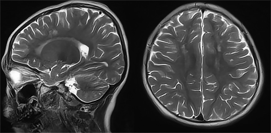
Multi-Organ MRI
MRI is another advanced imaging technique that provides detailed images of the body. It uses magnetic fields and radio waves to create detailed images of internal structures9. MRI machines contain large, powerful magnets that generate a strong magnetic field around the patient. When a patient is positioned inside the MRI machine, the magnetic field aligns the hydrogen atoms in the body in a specific direction.
The MRI machine then sends radio-frequency (RF) pulses, which temporarily alter the alignment of hydrogen atoms. When the RF pulses cease, the hydrogen atoms return to their original alignment, emitting energy as radio waves detected by the MRI scanner.
Due to their differing water contents, different tissues in the body have varying amounts of hydrogen atoms and thus release energy at different rates. The MRI scanner can differentiate between different tissue types and produce high-resolution cross-sectional images10.
With MRI, your radiologist can observe oedema11, inflammatory sites12,13, and other abnormalities, such as tumours and precancerous lesions14. An MRI scan is highly sensitive, fast, painless, and free of ionising radiation.
What To Expect During an MRI Scan
MRI uses a strong magnetic field. As such, you will be asked to leave metal objects like your watch, mobile phone, jewellery, glasses, and a belt outside the scanner room. Your safety is our priority, so if you have a pacemaker or any non-removable metal items inside your body, you should consult our team, who may be able to clear you for safety or recommend a more suitable alternative.
Once in the exam room, you will lie face up on the bed, which will then slide into the scanner. Since the scanner is tunnel-like, you should inform your doctor beforehand if you are claustrophobic so they can provide appropriate medication. The scanning process involves turning the magnet on and off for over 60 minutes.
At times, the technologist may instruct you to hold your breath to enhance image quality, as movement, including breathing, can affect the results. Once the scan is complete, the radiology staff will assist you in retrieving your belongings.
What You Need To Know About Multi-Organ-Body Scans
Keep the following in mind before your scan:
- CT scans use X-rays, a form of ionising radiation. Therefore, scans should be performed when necessary, such as in high-risk populations. Even for scans that use higher doses of ionising radiation, the risk of cancer is still low and is outweighed by the importance of getting the right diagnosis and treatment for at-risk individuals15.
- MRI scans are highly sensitive. However, with this comes a risk of overdiagnosis and overtreatment since not all pre-cancer or abnormalities will turn into cancer, especially in healthy people16.
- According to the FDA, the medical benefits of full-body CT scans have not yet been sufficiently proven, though evidence has shown targeted CT scans, such as lung CTs, to be highly effective.
- Routine LDCT scans can be particularly effective for people at high risk of lung cancer, reducing mortality by up to 25 percent in men and 61 percent in women5.
- Even if you choose to have a multi-organ scan, it is still recommended that you continue annual mammograms to screen for early-stage breast cancer or a colonoscopy to screen for early colon cancer signs.
Should You Get a Multi-Organ Scan?
You should make an informed decision about whether to receive regular multi-organ scans. To help you decide, consider your risk factors for developing pathologies such as cancer17.
You should consider getting an annual screening if you fit one or more of the following criteria:
- You have a family history of aneurysms.
- You are overweight.
- You are sedentary.
- You are 45+ years old.
- You have high cholesterol, diabetes, or high blood pressure.
- If you have smoked at least one pack of cigarettes per day for a minimum of 10 years, you should consider an add-on LDCT scan. Even if you do not smoke anymore, you still have a higher lung cancer risk.
Conclusion
In conclusion, multi-organ scans offer a comprehensive approach to proactive health management, enabling the early detection of potential health issues before symptoms arise. Utilising advanced imaging technologies such as CT and MRI, these scans provide detailed insights into your body's internal structures, helping to identify signs of conditions like cancer at treatable stages. By understanding what scans can reveal and considering your personal risk factors, you can make an informed decision about whether to incorporate annual screenings into your healthcare routine.
If you want to be proactive about your health, why not book Ezra's MRI scan or Lung CT scan? Our annual scan catches potential signs of cancer earlier, leveraging AI through the screening process to make it more efficient, affordable, and faster.
Understand your risk for cancer with our 5 minute quiz.
Our scan is designed to detect potential cancer early.
References
1. NHS screening. nhs.uk. April 9, 2018. Accessed October 28, 2025. https://www.nhs.uk/tests-and-treatments/nhs-screening/
2. CT scan. nhs.uk. October 18, 2017. Accessed October 28, 2025. https://www.nhs.uk/tests-and-treatments/ct-scan/
3. Advancements in Computed Tomography Technology. Imaging Technology News. November 1, 2023. Accessed October 28, 2025. http://www.itnonline.com/article/advancements-computed-tomography-technology
4. Patel PR, De Jesus O. CT Scan. In: StatPearls. StatPearls Publishing; 2025. Accessed October 28, 2025. http://www.ncbi.nlm.nih.gov/books/NBK567796/
5. England NHS. NHS England » Rolling out targeted lung health checks. January 18, 2024. Accessed October 28, 2025. https://www.england.nhs.uk/blog/rolling-out-targeted-lung-health-checks/
6. Chowdhury MM, Zieliński LP, Sun JJ, et al. Editor’s Choice – Calcification of Thoracic and Abdominal Aneurysms is Associated with Mortality and Morbidity. Eur J Vasc Endovasc Surg. 2018;55(1):101-108. doi:10.1016/j.ejvs.2017.11.007
7. Gore RM. Abdominal Roentgenography. In: Walker HK, Hall WD, Hurst JW, eds. Clinical Methods: The History, Physical, and Laboratory Examinations. 3rd ed. Butterworths; 1990. Accessed October 28, 2025. http://www.ncbi.nlm.nih.gov/books/NBK205/
8. CT colonography | Tests and scans | Cancer Research UK. Accessed October 28, 2025. https://www.cancerresearchuk.org/about-cancer/tests-and-scans/ct-colonography
9. MRI scan. nhs.uk. October 23, 2017. Accessed October 28, 2025. https://www.nhs.uk/tests-and-treatments/mri-scan/
10. Thomas KE, Fotaki A, Botnar RM, Ferreira VM. Imaging methods: magnetic resonance imaging. Circ Cardiovasc Imaging. 2023;16(1):e014068. doi:10.1161/CIRCIMAGING.122.014068
11. Lindow T, Quadrelli S, Ugander M. Noninvasive Imaging Methods for Quantification of Pulmonary Edema and Congestion. JACC Cardiovasc Imaging. 2023;16(11):1469-1484. doi:10.1016/j.jcmg.2023.06.023
12. Ferreira VM, Schulz-Menger J, Holmvang G, et al. Cardiovascular Magnetic Resonance in Nonischemic Myocardial Inflammation. JACC. 2018;72(24):3158-3176. doi:10.1016/j.jacc.2018.09.072
13. Association between knee inflammation and knee pain in patients with knee osteoarthritis: a systematic review - Osteoarthritis and Cartilage. Accessed October 28, 2025. https://www.oarsijournal.com/article/S1063-4584(21)01003-7/fulltext
14. MRI scan | Tests and scans | Cancer Research UK. Accessed October 28, 2025. https://www.cancerresearchuk.org/about-cancer/tests-and-scans/mri-scan
15. Can ionising radiation cause cancer? Cancer Research UK. March 24, 2015. Accessed October 28, 2025. https://www.cancerresearchuk.org/about-cancer/causes-of-cancer/air-pollution-radiation-and-cancer/ionising-radiation-and-cancer
16. Vickers A. Effects of Magnetic Resonance Imaging Targeting on Overdiagnosis and Overtreatment of Prostate Cancer. Eur Urol. 2021;80(5):567-572. doi:10.1016/j.eururo.2021.06.026
17. Cancer causes and risk factors explained. Accessed October 28, 2025. https://www.macmillan.org.uk/cancer-information-and-support/worried-about-cancer/causes-and-risk-factors


