Chronic obstructive pulmonary disease (COPD) is one of the leading causes of illness and disability in the UK, yet many people don’t realise they’re developing lung damage until it’s advanced. The early stages often go unnoticed, especially in those at higher risk, like smokers or people exposed to air pollutants. That’s where low-dose computed tomography (LDCT) scans come in. These scans can reveal subtle signs of lung damage early, supporting faster diagnosis and better long-term outcomes.
What is a Low-Dose CT Scan, and How Does it Work?
An LDCT is a type of medical imaging procedure that uses a computer linked to an X-ray machine to produce detailed cross-sectional pictures of the body’s internal organs, such as the lungs, but with a much lower dose of radiation than a standard CT scan1. This allows healthcare providers to monitor lung health and detect abnormalities while minimising the risk of potential radiation exposure from frequent or routine screenings.
CT imaging involves the patient lying on a table that moves through a circular scanner. The scanner rotates around the body, capturing numerous X-ray images from different angles2. A computer then processes these images to create detailed cross-sectional “slices” of the internal organs, such as the lungs, providing a highly detailed 3D view that helps identify and evaluate conditions like emphysema, cancer, and other lung diseases.
What Makes it Low-Dose?
An LDCT scan uses about one-fifth as much radiation as a standard CT scan. A standard chest CT involves roughly 7 mSv (millisieverts) of radiation, while an LDCT is closer to 1.4-2 mSv, equivalent to around six months of natural background radiation3.
Compared to a chest X-ray, which delivers 0.1-0.2 mSv, low-dose CT scans do involve slightly more radiation, but they achieve much higher image clarity and detailed cross-sectional views4. Advances in CT technology mean these scans maintain a high diagnostic quality, enabling early detection of small lung changes with far less exposure than standard CT techniques.
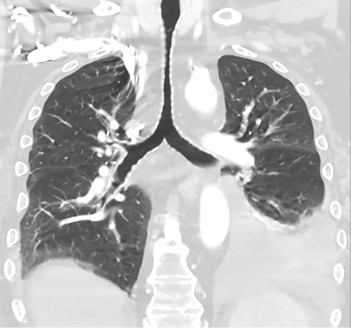
How LDCT Detects COPD and Lung Damage
LDCT can detect signs of lung damage, including COPD, by revealing key features such as over-inflated air spaces (areas of lung destruction characteristic of emphysema), thickened airway walls, and regions of scarring or fibrosis. Emphysema appears on CT as areas where the alveolar walls are destroyed, resulting in larger, abnormal air spaces and reduced lung density5. Chronic bronchitis is indicated by visible airway thickening and mucus accumulation in the bronchi6.
Spotting Early Structural Changes
LDCT scans can reveal subtle lung damage, such as early emphysema or airway abnormalities, even before noticeable symptoms appear7. This is particularly valuable for people with a history of smoking or occupational exposures, as early detection enables proactive management (like smoking cessation or other interventions) before the disease becomes advanced and symptomatic.
CT as Part of a Broader Diagnostic Pathway
While CT imaging can identify and map the extent of lung damage, it is typically used in combination with spirometry (which measures lung function) and thorough clinical assessments8. This integrated approach ensures that a diagnosis of COPD is both accurate and comprehensive, using imaging to confirm the presence, severity, and progression of disease picked up by clinical and functional testing. CT scans are especially helpful for monitoring change over time or assessing complex cases where function tests alone are inconclusive.
Benefits of Early Detection with CT Imaging
Early diagnosis enables earlier, more targeted treatment, often leading to better outcomes. When COPD is identified in its initial stages, clinicians can introduce interventions like smoking cessation support, pulmonary rehabilitation, and appropriate medications before irreversible damage occurs. These measures can help slow disease progression and preserve lung function for longer.
Reduced Hospitalisations and Improved Quality of Life
Ongoing CT monitoring and early diagnosis can make a significant impact in the UK. COPD is the second leading cause of emergency hospital admissions nationwide and accounts for around 1.4 million GP consultations every year9. Timely, quality-assured diagnosis, often aided by imaging, can help reduce harmful flare-ups and lower the heavy burden COPD places on the NHS by decreasing unplanned hospital admissions.
One 2021 study highlighted a 32 per cent reduction in COPD emergency admissions in the UK, from 239 per 100,000 people in 2019 to 163 per 100,000 in 2021, indicating that robust early intervention and monitoring strategies, including images, have real-world benefits9,10. This approach gives people with COPD a better chance to manage their symptoms and improve their well-being, while easing pressure on healthcare resources.
Detecting Other Lung Conditions
LDCT may also reveal early lung nodules or signs of lung cancer, often before symptoms arise. Comprehensive screening in high-risk individuals not only helps detect COPD and emphysema but can simultaneously identify cancers, infections, or other lung abnormalities, making it a valuable tool for broad lung health surveillance11,12.
Who Should Consider an LDCT Scan?
- Smokers or ex-smokers over age 45.
- Individuals with a family history of lung disease.
- People with significant exposure to air pollution, asbestos, or dust in the workplace or local environment.
Current NHS protocols and targeted lung health screening programmes focus particularly on adults between 55 and 74 with a clear history of tobacco use, but risk assessment also accounts for occupational and environmental exposures, as well as personal and family history of respiratory disease.
Asymptomatic Individuals with Risk Factors
Symptoms of COPD, such as breathlessness or chronic cough, often appear only after significant lung damage has already occurred13. This means many high-risk individuals may have early disease without realising it. Screening with LDCT enables the detection of structural lung changes before symptoms develop, supporting earlier treatment and better long-term health outcomes, even in those who feel well at the time of screening14.
Summary: LDCT and COPD
LDCT scans are a safe, highly effective tool for detecting COPD and lung damage at an early stage, using only a fraction of the radiation of standard CT while preserving high diagnostic accuracy. This enables clinicians to identify subtle airway changes or lung tissue damage long before symptoms escalate, ultimately empowering people to take charge of their lung health.
If you want to take charge of your lung health, why not book an Ezra Lungs CT Scan? For £249, you can catch potential abnormalities, including lung cancer, pulmonary nodules, emphysema, and more.
Understand your risk for cancer with our 5 minute quiz.
Our scan is designed to detect potential cancer early.
References
1. Definition of low-dose CT scan - NCI Dictionary of Cancer Terms - NCI. February 2, 2011. Accessed November 19, 2025. https://www.cancer.gov/publications/dictionaries/cancer-terms/def/low-dose-ct-scan
2. Computed Tomography (CT). National Institute of Biomedical Imaging and Bioengineering. Accessed November 19, 2025. https://www.nibib.nih.gov/science-education/science-topics/computed-tomography-ct
3. Radiology (ACR) RS of NA (RSNA) and AC of. Radiation Dose. Radiologyinfo.org. Accessed November 19, 2025. https://www.radiologyinfo.org/en/info/safety-xray
4. Wassipaul C, Janata-Schwatczek K, Domanovits H, et al. Ultra-low-dose CT vs. chest X-ray in non-traumatic emergency department patients – a prospective randomised crossover cohort trial. eClinicalMedicine. 2023;65. doi:10.1016/j.eclinm.2023.102267
5. Pahal P, Avula A, Afzal M. Emphysema. In: StatPearls. StatPearls Publishing; 2025. Accessed November 20, 2025. http://www.ncbi.nlm.nih.gov/books/NBK482217/
6. Widysanto A, Goldin J, Mathew G. Chronic Bronchitis. In: StatPearls. StatPearls Publishing; 2025. Accessed November 20, 2025. http://www.ncbi.nlm.nih.gov/books/NBK482437/
7. Steiger D, Siddiqi MF, Yip R, Yankelevitz DF, Henschke CI, I-ELCAP investigators. The importance of low-dose CT screening to identify emphysema in asymptomatic participants with and without a prior diagnosis of COPD. Clin Imaging. 2021;78:136-141. doi:10.1016/j.clinimag.2021.03.012
8. Chronic obstructive pulmonary disease (COPD) - Diagnosis. nhs.uk. October 20, 2017. Accessed November 20, 2025. https://www.nhs.uk/conditions/chronic-obstructive-pulmonary-disease-copd/diagnosis/
9. Potentially preventable emergency admissions. Nuffield Trust. Accessed November 20, 2025. https://www.nuffieldtrust.org.uk/resource/potentially-preventable-emergency-hospital-admissions
10. The prevalence of COPD in the UK paints a sad picture of inequalities. Accessed November 20, 2025. https://www.sanofi.co.uk/en/your-health/medicines/breathe-equal/the-prevalence-of-copd-in-the-uk-paints-a-sad-picture-of-inequalities
11. Gendarme S, Maitre B, Hanash S, Pairon JC, Canoui-Poitrine F, Chouaïd C. Beyond lung cancer screening, an opportunity for early detection of chronic obstructive pulmonary disease and cardiovascular diseases. JNCI Cancer Spectr. 2024;8(5):pkae082. doi:10.1093/jncics/pkae082
12. Mulshine JL, Pyenson B, Healton C, et al. Paradigm shift in early detection: Lung cancer screening to comprehensive CT screening. European Journal of Cancer. 2025;218:115264. doi:10.1016/j.ejca.2025.115264
13. COPD - Symptoms | NHLBI, NIH. October 4, 2024. Accessed November 20, 2025. https://www.nhlbi.nih.gov/health/copd/symptoms
14. Sekine Y, Katsura H, Koh E, Hiroshima K, Fujisawa T. Early detection of COPD is important for lung cancer surveillance. European Respiratory Journal. 2012;39(5):1230-1240. doi:10.1183/09031936.00126011


