Conditions our MRI scans have found
Bilateral cervical ribs
A cervical rib is an extra rib that is present at birth and forms above the first (top-most) rib, growing from the base of the neck just above the collarbone. It may grow on one side or both (bilateral) and may reach down to attach to the first rib (a fully formed bony rib) or may not be fully formed (a thin strand of tissue fibers). These are usually asymptomatic (do not cause symptoms) but can occasionally be a cause of thoracic outlet syndrome (a group of disorders that occur when blood vessels or nerves are compressed in the space between the collarbone and the first rib). Symptoms of thoracic outlet syndrome include pain in the shoulders and neck, numbness, weakness, and coldness in the fingers.
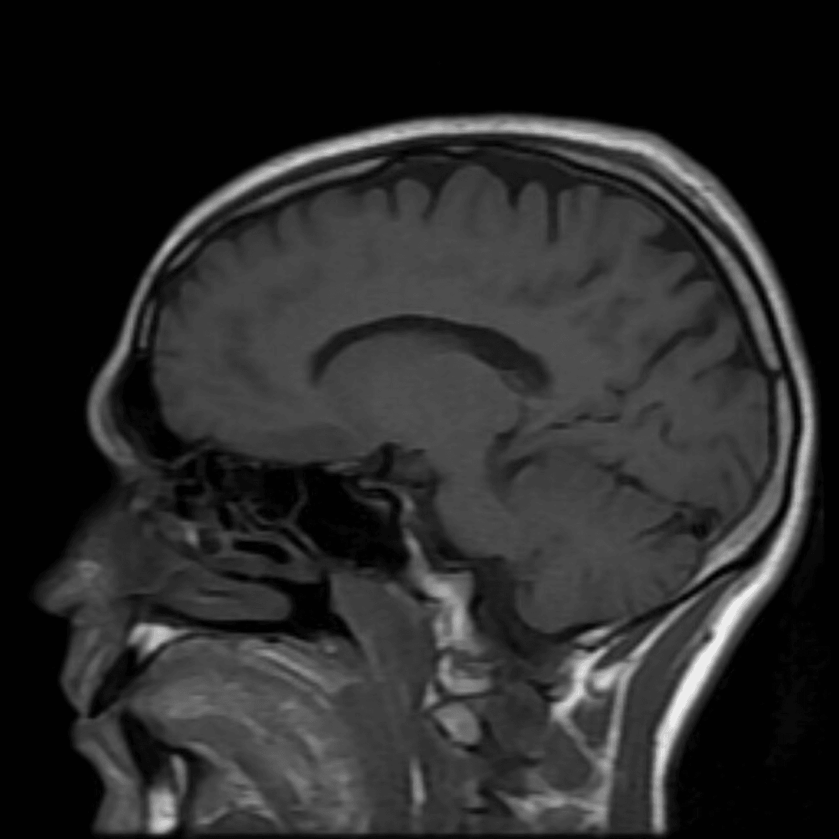
Bifrontal volume loss
Brain tissue tends to shrink at the rate of about 0.2% per year from the age of 30 and then accelerates after the age of 60 due to genetic, environmental, and lifestyle factors. Bifrontal volume loss sometimes can be associated with risk of dementia. There are currently no established guidelines for investigating or monitoring this condition.
Calcified lymph node
Lymph nodes are small bean-shaped structures that are part of the body's immune system. Lymph nodes filter substances that travel through the lymphatic fluid, and they contain lymphocytes (white blood cells) that help the body fight infection and disease. There are hundreds of lymph nodes found throughout the body. Calcium has a tendency to collect in healing tissue. Calcified structures in the body have no metabolic activity.
Bochdalek hernia
A Bochdalek hernia results when there is failure of closure of the diaphragm (a thin skeletal muscle that sits at the base of the chest and separates the abdomen from the chest) during embryonic development. This can allow protrusion of abdominal content, usually fat but sometimes organs or the intestines, into the chest. Bochdalek hernias are found in approximately 6% of adults, with incidence increasing with age. This is usually asymptomatic (does not cause symptoms), but can sometimes cause gastrointestinal (e.g. constipation, abdominal pain) or pulmonary symptoms.
Bladder wall lipoma
A bladder wall lipoma is a non-cancerous growth (i.e. tumor) of fat cells arising from the wall of the urinary bladder. Although lipomas are the most common non-cancerous masses found in the body, bladder tissue involvement is rare. This condition is typically found in passing (incidentally) on bladder imaging of individuals who are experiencing lower urinary tract symptoms (e.g. urgency, frequency, painful urination or incontinence) and/or the presence of blood in the urine. However, some individuals with a bladder wall lipoma may be asymptomatic (do not have symptoms). Although bladder wall lipomas do not carry any malignant (cancerous) potential, it must be differentiated from lesions that are potentially cancerous (e.g. liposarcoma).
Calcification of the prostate
Prostate calcifications (deposits of calcium in prostate tissue) are commonly seen in men as they age. Although the exact cause is not clearly understood, experts suggest the calcifications occur as a result of chronic inflammation of the prostate. Other potential causes include diabetes, infection, benign prostatic hypertrophy (BPH), radiation therapy, previous urethral stent placement/surgery, and prostate cancer.
Can't find what you're looking for?
Frequently Asked Questions
Unfortunately at this time, we are unable to scan people with pacemakers. There is a risk that the MRI magnetic fields will disrupt its operation, and we don't want that.
There are many different types of implants. We will need to know more about what you have. The good news is that almost all implants are MRI-safe. There is a chance the implant will affect the images we can get from the surrounding tissues.
Yes. Almost all IUDs are MRI-safe. Regardless, we check the MRI safety of all devices. Common MRI-safe IUDs are the Mirena and the Copper T.
Yes you can. There will be extra precautions we will take to ensure your safety while in the machine, so please inform staff before entering the MRI.
Yes. This is completely safe.