Conditions our MRI scans have found
Prostate volume
A normal prostate volume is approximately 20 cc. An enlarged prostate is >20 cc and may or may not cause urinary symptoms. If it does not cause any symptoms, then it does not need to be treated. The prostate volume is calculated based on a radiologist’s measurements. Enlarged prostate is not life threatening.
Prostate lesion
A prostate lesion is an area of tissue that has been damaged by injury or disease within the prostate. Prostate lesions could be benign (non-cancerous) or malignant (cancerous).
Prostate lesion with PI-RADS Score (1-5)
Radiologists use the Prostate Imaging Reporting and Data System (PI-RADS) score to report how likely it is that a suspicious area of the prostate is a clinically significant cancer. PI-RADS scores range from 1 (most likely not cancer) to 5 (very suspicious). Usually a score of 5 represents a high possibility that prostate cancer is present.
Proteinaceous kidney cyst
Cysts are fluid-filled sacs or pockets that can form in various parts of the body. Most cysts present little or no discomfort and are harmless. The majority disappear without treatment within a few months. Sometimes these cysts can contain thicker fluid, known as proteinaceous material.
PSA density
The PSA (prostate specific antigen) density is an additional tool that may be used to predict the likelihood of prostate cancer. It is the PSA level corrected for the size of the prostate. The PSA density is calculated by dividing the PSA level by the volume of the prostate. Prostate volume is calculated from MRI or transrectal ultrasound measurements. PSA Level ÷ Prostate Volume = Your PSA Density (ng/mL)Generally, if the PSA density is less than 0.15, a prostate biopsy can be reasonably avoided or delayed. A PSA density greater than or equal to 0.20 increases the suspicion of a clinically significant prostate malignancy (cancer).
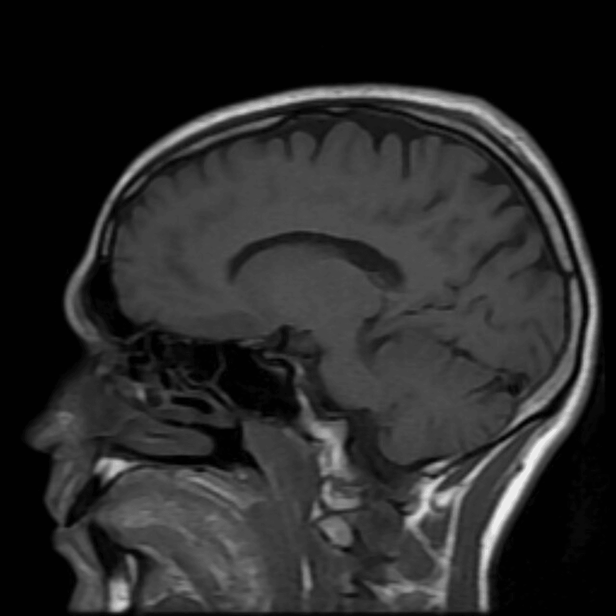
Prominent Meckel's
The brain and spinal cord are covered by three protective membrane-linings called meninges. The dura mater is the tough outermost layer. Meckel’s cave is a dura mater pouch filled with cerebral spinal fluid (CSF) at the base of the skull that contains the root of the trigeminal nerve (responsible for sensation of the face and movement in the jaw muscles). A prominent (enlarged) Meckel’s cave is a nonspecific finding (meaning it is difficult to say what the cause is).
Can't find what you're looking for?
Frequently Asked Questions
Unfortunately at this time, we are unable to scan people with pacemakers. There is a risk that the MRI magnetic fields will disrupt its operation, and we don't want that.
There are many different types of implants. We will need to know more about what you have. The good news is that almost all implants are MRI-safe. There is a chance the implant will affect the images we can get from the surrounding tissues.
Yes. Almost all IUDs are MRI-safe. Regardless, we check the MRI safety of all devices. Common MRI-safe IUDs are the Mirena and the Copper T.
Yes you can. There will be extra precautions we will take to ensure your safety while in the machine, so please inform staff before entering the MRI.
Yes. This is completely safe.