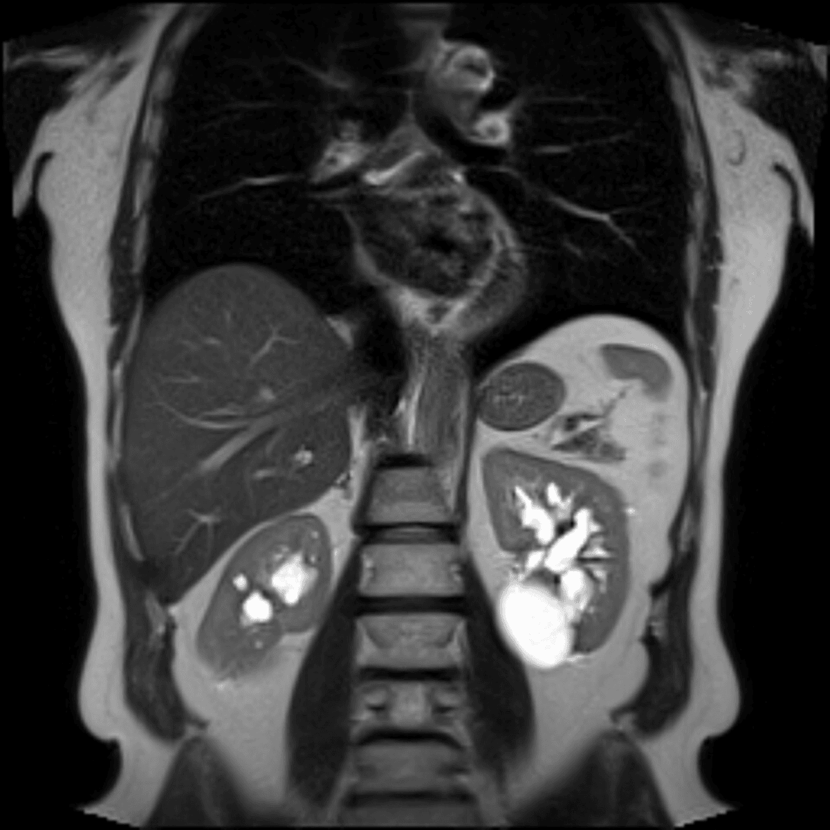
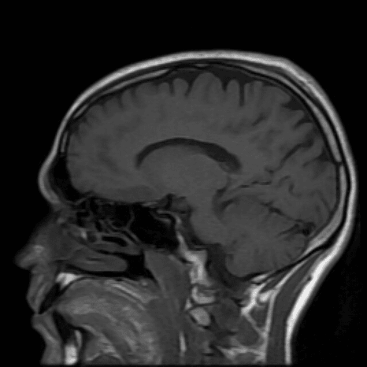
Pectus excavatum is when the chest wall is depressed (caved in); this is a congenital condition (from birth) that does not change much after puberty (when the bones have matured and hardened). Although the cause is unknown, it accounts for 90% of chest wall disorders. It is usually solely a cosmetic concern, but when it is clinically significant, it can cause decreased exercise tolerance or shortness of breath.
Phrygian cap is the most common congenital (present at birth) anatomical variant of the gallbladder. The gallbladder is divided into 3 parts - the neck, body, and fundus. A phrygian cap is when the fundus folds back onto the gallbladder body. This occurs in approximately 4% of individuals and is a benign (non-cancerous and of no clinical significance) finding.
Pelvic organ prolapse describes when pelvic organs (e.g. bladder, uterus, rectum) slip down from their normal anatomical position and push against or into the vagina. This is found in 3-11% of women, and is associated with having had multiple pregnancies, aging and obesity. Pelvic organ prolapse can cause symptoms including pelvic pressure or seeing tissue bulging out of the vagina.
Varices are caused by venous obstruction, increased pressure, and venous insufficiency, resulting in dilated (enlarged) and/or misshapen veins. Pregnancy, hormonal and anatomic factors play a role in their development. They can arise in isolation, in association with leg/buttock, vulvar, or labial varices, or as part of pelvic congestion syndrome (a condition that causes chronic pelvic pain).Symptoms can include pelvic pain, heaviness, and pressure that are exacerbated by prolonged standing, exercise, and coitus. Generally, treatment is only needed if symptoms are present.
A pelvic kidney is when one or both kidneys stay in the pelvis and do not move into their proper position during fetal development. A normal variant is an atypical finding that is seen but has no clinical significance, and is considered within the spectrum of normal findings. No further follow-up or evaluation is needed for this finding.
Peripelvic renal cysts are common findings of small fluid sacs within the renal pelvis (the enlarged upper end of the ureter, which carries urine from the kidney to the urinary bladder). Peripelvic cysts usually do not cause symptoms, but sometimes may grow in size leading to kidney infection (pyelonephritis), kidney stones, hematuria (blood in the urine) or hydronephrosis (swelling of a kidney due to a build-up of urine).