MRI for Kidney Cancer Detection
Kidney MRI, also known as renal MRI, utilises powerful magnets, typically with a strength of 1.5 or 3 Tesla, to generate detailed, cross-sectional images of the kidneys, surrounding fat, blood vessels, and the urinary tract. This technique provides exceptional soft-tissue contrast and structural information, making it valuable for both anatomical and functional assessment1,2.
Unlike CT scans, MRI does not use ionising radiation and offers several protocols tailored to patient needs. Multiphasic contrast MRI utilises gadolinium injection to visualise how a tumour enhances or “lights up” helping differentiate benign from malignant lesions3,4. For patients with kidney dysfunction or contrast allergies, non-contrast MRI can be used with core sequences (such as T1, T2, diffusion-weighted, and chemical-shift imaging) without the need for any injection1,5.
Learn more about MRI for cancer detection here.
Why You Might Need a Kidney MRI
There are several reasons why a kidney MRI might be ordered, including:
Blood in Urine with Inconclusive Ultrasound or CT
If you have blood in your urine (hematuria) but initial tests, such as ultrasound or CT scans, are unclear or inconclusive, a kidney MRI may be ordered. MRI offers clearer visualisation of the urinary tract and renal tissue, helping to detect tumours, cysts, or subtle structural changes that could be causing the bleeding6,7.
Investigating an Incidental Renal Mass
When an unexpected kidney mass is found on a routine scan, an MRI is used to characterise it, such as distinguishing a benign cyst from an angiomyolipoma (AML) or cancer6,7. Its superior soft-tissue contrast helps refine diagnosis and avoid unnecessary surgeries.
Staging Known Kidney Cancer
In patients already diagnosed with kidney cancer, MRI maps the extent of the tumour, including fat invasion, renal vein and inferior vena cava (IVC) thrombus, lymph node enlargement, and adjacent organ invasion. This precision helps guide surgical and treatment planning6,8.
Radiation-Free Surveillance for Hereditary Syndromes
People with certain hereditary risk factors, such as von Hippel-Lindau syndrome, require lifelong kidney surveillance9. MRI is preferred because it avoids radiation, minimises cumulative risk, and sensitively detects small tumours, cysts, or early-stage cancers that require close monitoring6.
For Patients with Poor Kidney Function or Gadolinium Allergy
If you have an eGFR below 30 or a history of allergy to iodine-based (CT) or gadolinium-based (MRI) contrast agents, a non-contrast kidney MRI is a safer alternative10,11. Non-contrast MRI provides critical diagnostic information without risking further kidney damage or allergic reactions.
Pregnancy: Safe, Detailed Imaging Without Radiation
During pregnancy, non-contrast MRI provides a detailed anatomical assessment of the kidneys, urinary tract, and surrounding tissues without the use of ionising radiation or contrast agents. This makes it the safest option when a detailed kidney evaluation is needed for expectant mothers12.
You can use our Know Your Risk Calculator to understand your risk of cancer in just five minutes.
How to Prepare for Your Kidney MRI
Here are a few tips to help you prepare for your MRI13:
- Take your usual medications and eat normally, unless instructed to fast for specific MRI protocols. Fasting is not always required however14.
- Avoid heavy caffeine intake and stay hydrated.
- Undergo a recent blood creatine/eGFR test (within 30 days) to confirm it is safe to receive gadolinium.
- Inform staff about any metal implants or devices, and bring safety cards for “MR-Conditional” implants, such as pacemakers or aneurysm clips.
- Remove all metal items, including jewellery, hairpins, dental plates, and transdermal patches with foil, to prevent image distortion.
- Bring your referral, prior brain images, and insurance pre-authorisation to avoid delays.
- Wear comfortable, metal-free clothing; ask about earplugs, music, or mirror goggles if you’re claustrophobic.
- If contrast is planned, fast for 4–6 hours prior to the procedure. Inform staff about any kidney issues or past reactions to contrast agents, as safer alternatives may be available.
- Arrange an escort home if you need sedation for claustrophobia.
You can read more about preparation for Ezra’s Full Body Scan here.
What Happens During the Kidney Scan?
Upon arrival for your MRI, you will need to check in and complete a screening form. This will allow you to confirm the presence of implants, allergies, and whether you might need any anxiety medication.
A kidney MRI scan is a detailed, non-invasive imaging procedure that can be performed with or without the use of contrast. Here’s what you can expect during the scan:
Common Steps (Both Contrast and Non-Contrast Protocols)
- Patient Positioning: You’ll lie on your back (supine), entering the MRI machine feet-first. A specialised torso surface coil is placed over your abdomen to optimise image quality.
- Initial Sequences: The scan begins with a fast T2-weighted sequence (HASTE or FRFSE), which excels at revealing fluid-filled areas, cysts, and signs of hydronephrosis (swelling of the kidney).
- Anatomic Imaging: Both axial (cross-sectional) and coronal (front-to-back) T1-weighted images are obtained in in-phase and out-of-phase modes. These sequences are particularly effective in detecting fat, internal bleeding, or protein-rich cysts within the kidneys.
- Functional Assessment: Diffusion-weighted imaging is performed, and an ADC (apparent diffusion coefficient) map is generated. This step helps identify highly cellular or aggressive tumours by observing how water molecules move through tissues15.
Contrast Pathway
If a contrast-enhanced study is needed16:
- Contrast Injection: An intravenous bolus of gadolinium-based contrast is administered to highlight vascular structures and enhance tissue detail.
- Corticomedullary Phase (≈ 25 seconds): Early arterial imaging helps visualise tumour “blush” and differentiate vascular from non-vascular tissues.
- Nephrographic Phase (≈ 80 seconds): Provides optimal visualisation of solid kidney masses against background renal tissue.
- Delayed/Urographic Phase (3–10 minutes): Late imaging highlights the lining of the urinary tract (urothelium) and lymphatic drainage; this phase is crucial for assessing urothelial involvement or abnormalities in the collecting system.
- Optional Vascular Imaging: Techniques such as TWIST MRA or dynamic contrast-enhanced (DCE) perfusion provide additional detail about blood vessels or perfusion patterns when needed.
Non-Contrast Pathway
If no intravenous contrast is given:
- Omitting the IV Injection: The scan proceeds without the administration of gadolinium.
- Chemical-Shift Imaging: Additional chemical-shift pairs are acquired to better identify intracellular fat, a feature of certain benign or malignant lesions.
- Advanced Research Options: Optional sequences like BOLD (blood oxygen level-dependent) imaging or arterial spin labelling (ASL) perfusion offer information about tissue oxygenation and blood flow, all without using contrast agents.
Typical Table Time: Expect to spend around 10–15 minutes in the scanner for a non-contrast study and about 25 minutes if contrast is used, depending on the protocol specifics and need for extra imaging.
At Ezra, our Full Body Plus scan takes around 60 minutes total, with 45 minutes of table time. Earplugs or headphones are available.
MRI Safety, Risks & Side-Effects
MRI is generally considered very safe when proper screening and protocols are followed, but certain risks and side effects should be understood:
- Metal and implants: The strong 3-Tesla magnet can pull or heat older pacemakers, aneurysm clips, or metal fragments17. Most modern “MR-Conditional” devices (like cochlear implants or pain pumps) are safe after screening, but all implants must be checked before scanning18.
- Gadolinium contrast: Macrocyclic gadolinium agents (e.g., gadobutrol) have an extremely low risk of allergic reactions or nephrogenic systemic fibrosis (NSF) when kidneys are healthy19. However, gadolinium can accumulate in tissues, and rare side effects such as headaches or skin changes have been reported. Many centres now offer contrast-free alternatives for routine follow-up20. You can read more about gadolinium contrast side effects here.
- Incidental findings: Focal liver lesions are the most frequent incidental findings on MRI, seen in up to 5-18% of imaging studies, with the majority representing benign cysts, hemangiomas, or focal nodular hyperplasia21–23.
- Claustrophobia: Anxiety inside the scanner is common. Wide-bore scanners, music, mirror goggles, or a single dose of oral sedative can help alleviate symptoms. Open MRI is an option if image detail can be sacrificed.
- Zero Ionising Radiation: MRI uses magnetic fields and radio waves, not X-rays, so there is no ionising radiation exposure, making it safer for repeated scans compared to CT scans24.
- Minor Sensations: Expect loud knocking, mild table vibration, and a brief cool flush if contrast is injected. Rare side effects include headaches, fatigue, or mild skin heating.
A deeper dive into possible side effects (such as heat, headaches, and gadolinium deposition) is available in our full guide.
At Ezra, we employ a contrast-free approach using wide-bore T3 machines to deliver a comfortable scanning experience.
Terms You Might See in Your MRI Report (And What They Might Mean)
MRI reports for kidney scans often include specialised terms. Here’s a guide to some common phrases and what they mean for your diagnosis:
- Corticomedullary / Nephrographic / Excretory phase: These terms describe specific timings of post-contrast MRI images—corticomedullary phase (early, highlights vessels), nephrographic phase (intermediate, best for tumour detection), and excretory phase (late, visualises the collecting system and ureters)25,26.
- Hyper- vs. hypo-enhancing mass: A hyper-enhancing mass appears brighter than the normal kidney after contrast, often indicating a highly vascular tumour such as clear-cell RCC; a hypo-enhancing mass appears darker and is less vascular or possibly benign27.
- Pseudocapsule sign: This is a thin, dark rim seen around a tumour, commonly associated with clear cell renal cell carcinoma and can help guide surgical planning28.
- ADC value: The apparent diffusion coefficient is a number from diffusion-weighted imaging; lower values mean the tumour is more cellular and likely malignant, while higher values suggest benignity29.
- Bosniak III/IV cyst: Complex renal cysts classified as Bosniak III (indeterminate but suspicious, 40–60% cancer risk) or Bosniak IV (highly suspicious, >80% risk); these often require surgical evaluation or removal30,31.
- Perinephric fat stranding/invasion: Refers to streaky changes or direct tumour extension into the fat surrounding the kidney, indicating that the tumour has spread beyond the capsule (T3a disease)32.
- Tumour-thrombus Level I–IV: Describes how far a tumour-related clot (thrombus) extends into the renal vein or inferior vena cava, with higher levels indicating further extension and more complex treatment requirements33.
- Fat-suppressed T1: An MRI sequence where fatty tissue appears dark, used to highlight fatty tumours like angiomyolipoma (AML); macroscopic fat becomes obvious on these images34.
Ezra provides a radiologist-reviewed report in a non-technical and easy-to-understand format on your dashboard.
After the MRI Scan
After the MRI scan, you will be free to go home and continue with your day without any precautions35. If you received a sedative, you will need another person to pick you up. You will also not be able to drive, consume alcohol or operate heavy machinery 24 hours after the sedative.
A team of experts will review your results and determine whether a follow-up is necessary and recommend the appropriate treatment if needed. If abnormalities are found, you may undergo ongoing monitoring every 2-3 months to track recurrence. You can receive support in the form of counselling and advice on how to handle aspects like claustrophobia.
If you have a scan with us here at Ezra, you will receive your report within five to seven days and have the option to discuss it with a medical practitioner. You can also access your scan images through the online portal.
What MRI Can Show About Kidney Cancer
Exact Size and Location of a Mass
MRI precisely measures the tumour’s dimensions and shows its location in relation to the renal hilum (the area where blood vessels, nerves and the ureter connect to the kidney) and nearby vessels36. This spatial accuracy is crucial for determining surgical or interventional strategies.
Enhancement Pattern Suggesting Subtype
The way a tumour “lights up” after contrast (enhancement pattern) can hint at its subtype. For instance, clear-cell renal cell carcinomas (RCCs) often exhibit strong, early enhancement, which helps differentiate them from other types, such as papillary or chromophobe RCC37.
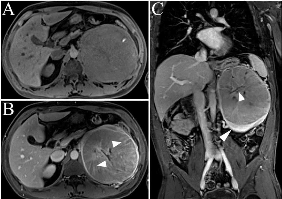
Pseudocapsule Sign
Many clear-cell RCCs are surrounded by a thin, dark rim (the pseudocapsule) on MRI. Recognising this sign supports the diagnosis of clear-cell RCC and can guide surgeons toward nephron-sparing procedures38.
Perinephric Fat Invasion
MRI can show the loss of the normal bright fat plane around the kidney, suggesting that the tumour has invaded the surrounding (perinephric) fat36. This finding upstages the disease to at least T3a and has important implications for surgical planning and prognosis.
Tumour-Thrombus Tracking
MRI excels at following tumour-related clot (tumour-thrombus) extending from the renal vein into larger vessels, such as the inferior vena cava (IVC) and even the right atrium of the heart39. Visualising the entire course of such a thrombus is critical for operative strategy and risk assessment.
Regional Lymph Node and Adrenal Involvement
In a single scan, MRI can also evaluate the lymph nodes and adrenal glands near the kidney. Detecting enlarged or abnormal nodes, or adrenal invasion, influences both staging and the extent of surgery required40.
Adjacent Organ Encroachment
MRI assesses whether the tumour has come into contact with or invaded organs near the kidney, such as the psoas muscle, colon, or liver41. Involvement of these structures can alter surgical plans, potentially requiring more extensive or multidisciplinary intervention.
Ezra utilises DWI as part of our whole-body MRI scans and artificial intelligence (AI) to enhance MRI images and convert radiology reports into layman's term translations.
Types of Kidney Tumours and How They Look on MRI
Clear-cell renal cell carcinoma (RCC) typically appears very bright on T2-weighted MRI, showing strong, often heterogeneous arterial enhancement after contrast administration42. It also displays low ADC values on diffusion sequences, reflecting its high cellularity. A thin, dark pseudocapsule may also be visible, aiding in the distinction of this aggressive subtype43.
Papillary RCC is characteristically dark on T2-weighted images, demonstrates weak or slow progressive enhancement with gadolinium, and may show high signal intensity on in-phase T1 due to hemosiderin, making it less vascular and distinct from clear-cell RCC44.
Chromophobe RCC shows intermediate signal intensity on T2-weighted MRI, tends to be even in appearance, and enhances moderately and evenly with contrast, generally lacking the strong “wash-in” seen in clear-cell RCC45.
Oncocytoma is a benign tumour that can mimic chromophobe RCC on MRI by showing homogenous enhancement and intermediate to high T2 signal; sometimes, a central stellate scar is present, which is suggestive but not exclusive to oncocytoma46.
Angiomyolipoma (AML) is identified by signal dropout of macroscopic fat on fat-suppressed T1-weighted images, while classic AMLs appear bright on T2, but fat-poor or hemorrhagic AMLs lack visible fat and may resemble malignant masses47.
Cystic lesions classified as Bosniak II-IV are characterised by complex internal structures, including septations, mural nodules, or irregular walls48. MRI allows for the confident detection of contrast wash-in or enhancement on subtraction images, with Bosniak III/IV cysts carrying a higher malignancy risk.
Wilms tumour, a childhood renal cancer, appears as a large heterogeneous mass that frequently crosses the midline and may displace adjacent organs; MRI is especially useful for follow-up to avoid radiation exposure and can show tumour components such as necrosis or haemorrhage49.
Upper-tract urothelial carcinoma is typically characterised by focal wall thickening or a mass affecting the renal pelvis or ureter, most visible during the delayed (excretory) phase of imaging. MRI can help define the tumour extent and guide surgical management50.
Ezra screens for over 500 conditions, including the kidneys.
Types of MRI Scans Used in Kidney Cancer Detection
There are multiple types of MRI scans, all using different methods to give a better visualisation of kidney tumours.
- Conventional 1.5 Tesla MRI is the most widely used system for renal imaging. It offers a pragmatic balance between spatial resolution and resistance to imaging artefacts, making it a mainstay for routine kidney cancer evaluations2.
- 3 Tesla MRI systems enable higher signal-to-noise ratios and facilitate the acquisition of thinner slices, which can improve the detection of small tumours, especially those less than 1 cm in size51. Multiparametric protocols involve various sequences (T1, T2, DWI, and contrast-enhanced phases) to distinguish tumour subtypes and differentiate benign from malignant lesions52.
- DCE MRI involves rapid imaging during the arterial, nephrographic, and delayed post-contrast phases53. This technique provides detailed enhancement curves, allowing for the assessment of tumour vascularity and permeability. Key quantitative parameters, such as Ktrans, can serve as biomarkers for evaluating tumour aggressiveness and response to anti-angiogenic therapies54.
- DWI evaluates the movement of water molecules in tissue and is highly sensitive to tumour cellularity55. Malignant tumours, which are densely cellular, typically demonstrate restricted diffusion and consequently lower ADC values56. ADC mapping helps differentiate benign from malignant renal masses and can stratify RCC subtypes.
- Time-resolved MR angiography techniques such as TWIST (Time-resolved Angiography with Interleaved Stochastic Trajectories) capture the dynamics of blood flow in the renal arteries and veins57. This is particularly valuable for visualising tumour thrombus extension in renal veins or the inferior vena cava, and for evaluating complex vascular anatomy before surgery.
- Blood-Oxygen-Level-Dependent (BOLD) and Arterial Spin Labelling (ASL) MRI are advanced research tools exploring tissue oxygenation and blood flow, respectively. BOLD detects hypoxic regions within tumours, while ASL measures quantitative perfusion without the use of contrast agents, making it valuable when gadolinium is contraindicated. Both techniques are promising for assessing tumour physiology, therapy response, and transplant function, but their clinical application remains investigational58.
- MR elastography is an emerging modality that measures tissue stiffness. Tumours may show distinct stiffness profiles compared to surrounding renal parenchyma or fibrotic tissues, offering a potential non-invasive biomarker for distinguishing malignant lesions from benign fibrotic processes59. While promising, MR elastography is largely experimental in the routine assessment of kidney cancer60.
Ezra uses whole-body DWI imaging to get a full picture of the body and catch any potential abnormalities.
MRI vs. Other Imaging Tests for Kidney Cancer Detection
MRI Scan Cost
Ezra’s MRI Scan with Spine costs £2,395 and is currently available at their partner clinic in Marylebone, London and in Sidcup, with more locations planned in the future. No referral is required, so you can book your scan directly without first consulting a GP or specialist. Most people pay out-of-pocket, as insurance typically does not cover self-referred scans, but you may be able to seek reimbursement depending on your policy.
Frequently Asked Questions
Does your whole body go in for a kidney MRI?
For a dedicated kidney MRI, only the part of your body being scanned, typically your abdomen, is placed inside the scanner, not your entire body.
Which is better, an MRI or a CT scan for the kidneys?
CT scans are generally superior for detecting kidney stones and calcifications, while MRI provides better soft tissue contrast for tumours and is used when avoiding radiation is important.
Will an MRI show a kidney stone?
MRI can sometimes detect kidney stones, but it is much less accurate than CT, which remains the gold standard for identifying stones.
Key Takeaways
- Kidney MRI, with or without contrast, provides the most detailed, radiation-free assessment for detecting, sizing, and staging renal tumours less than 4 cm, making early nephron-sparing surgery possible.
- If gadolinium is safe, contrast-enhanced MRI improves the accuracy of subtype identification, Bosniak cyst classification, and surgical planning.
- When gadolinium is not safe, non-contrast MRI, which utilises diffusion and chemical-shift techniques, still surpasses ultrasound and often rivals CT in detecting solid masses, all without the use of nephrotoxic dye.
- High-risk patients, such as those with a history of smoking, hypertension, or familial syndromes, benefit from regular MRI surveillance, as early detection of RCC leads to higher cure rates, smaller surgeries, and better long-term kidney function.


References
1. Francis ST, Selby NM, Taal MW. Magnetic Resonance Imaging to Evaluate Kidney Structure, Function, and Pathology: Moving Toward Clinical Application. American Journal of Kidney Diseases. 2023;82(4):491-504. doi:10.1053/j.ajkd.2023.02.007
2. Nikken JJ, Krestin GP. MRI of the kidney—state of the art. Eur Radiol. 2007;17(11):2780-2793. doi:10.1007/s00330-007-0701-3
3. Bodard S, Cornelis FH. Non-invasive functional MRI techniques for early detection of kidney injury in chronic kidney disease: a positive step forward. Annals of Translational Medicine. 2024;12(4):80-80. doi:10.21037/atm-23-1788
4. Schieda N, Krishna S, Davenport MS. Update on Gadolinium-Based Contrast Agent–Enhanced Imaging in the Genitourinary System. American Journal of Roentgenology. 2019;212(6):1223-1233. doi:10.2214/AJR.19.21137
5. 3T MR imaging protocol for characterization of renal masses | Applied Radiology. Accessed July 17, 2025. https://appliedradiology.com/articles/3t-mr-imaging-protocol-for-characterization-of-renal-masses
6. Willatt J, Francis IR. Imaging and management of the incidentally discovered renal mass. Cancer Imaging. 2009;9(Special issue A):S30-S37. doi:10.1102/1470-7330.2009.9008
7. Agnello F, Albano D, Micci G, et al. CT and MR imaging of cystic renal lesions. Insights Imaging. 2020;11:5. doi:10.1186/s13244-019-0826-3
8. Reznek RH. CT/MRI in staging renal cell carcinoma. Cancer Imaging. 2004;4(Spec No A):S25-S32. doi:10.1102/1470-7330.2004.0012
9. Ashouri K, Mohseni S, Tourtelot J, et al. Implications of Von Hippel-Lindau Syndrome and Renal Cell Carcinoma. J Kidney Cancer VHL. 2015;2(4):163-173. doi:10.15586/jkcvhl.2015.41
10. Gadolinium contrast injection - Overview. Guy’s and St Thomas’ NHS Foundation Trust. Accessed July 17, 2025. https://www.guysandstthomas.nhs.uk/health-information/gadolinium-contrast-injection
11. Martino F, Amici G, Rosner M, et al. Gadolinium-Based Contrast Media Nephrotoxicity in Kidney Impairment: The Physio-Pathological Conditions for the Perfect Murder. J Clin Med. 2021;10(2):271. doi:10.3390/jcm10020271
12. Gatta G, Di Grezia G, Cuccurullo V, et al. MRI in Pregnancy and Precision Medicine: A Review from Literature. J Pers Med. 2021;12(1):9. doi:10.3390/jpm12010009
13. Radiology (ACR) RS of NA (RSNA) and AC of. Magnetic Resonance Imaging (MRI) - Head. Radiologyinfo.org. Accessed July 3, 2025. https://www.radiologyinfo.org/en/info/mri-brain
14. Williams JM, Hilmes MA, Archer B, et al. Repeatability and Reproducibility of Pancreas Volume Measurements Using MRI. Sci Rep. 2020;10:4767. doi:10.1038/s41598-020-61759-9
15. Kitazume Y, Satoh S, Taura S, et al. Diffusion-weighted magnetic resonance imaging detection of renal cancer presenting with diffuse peritoneal metastases in a patient with hemodialysis-associated acquired cystic disease of the kidney. J Magn Reson Imaging. 2009;29(4):953-956. doi:10.1002/jmri.21640
16. Thorson D, Bova D, Picken MM, et al. Peak early‐phase enhancement ratio on contrast‐enhanced MRI to differentiate chromophobe renal cell carcinoma from oncocytoma. BJUI Compass. 2025;6(4):e70017. doi:10.1002/bco2.70017
17. Gill A, Shellock FG. Assessment of MRI issues at 3-Tesla for metallic surgical implants: findings applied to 61 additional skin closure staples and vessel ligation clips. J Cardiovasc Magn Reson. 2012;14(1):3. doi:10.1186/1532-429X-14-3
18. Potential Hazards and Risks. UCSF Radiology. January 20, 2016. Accessed March 14, 2025. https://radiology.ucsf.edu/patient-care/patient-safety/mri/potential-hazards-risks
19. Costello JR, Kalb B, Martin DR. Incidence and Risk Factors for Gadolinium-Based Contrast Agent Immediate Reactions. Top Magn Reson Imaging. 2016;25(6):257-263. doi:10.1097/RMR.0000000000000109
20. McDonald RJ, McDonald JS, Kallmes DF, et al. Gadolinium Deposition in Human Brain Tissues after Contrast-enhanced MR Imaging in Adult Patients without Intracranial Abnormalities. Radiology. 2017;285(2):546-554. doi:10.1148/radiol.2017161595
21. Blum SFU, Ittermann T, Kromrey ML, et al. Long-term outcome of incidental cystic liver tumors in the general population. Sci Rep. 2021;11(1):11661. doi:10.1038/s41598-021-91140-3
22. Moreira-Silva H, Amorim J, Santos-Silva E. Incidental Liver Lesions in children: A practical and evidence-based approach. Clinics and Research in Hepatology and Gastroenterology. 2022;46(5):101904. doi:10.1016/j.clinre.2022.101904
23. Sawatzki M, Husarik DB, Semela D. Assessment of focal liver lesions in non-cirrhotic liver – expert opinion statement by the Swiss Association for the Study of the Liver and the Swiss Society of Gastroenterology. Swiss Medical Weekly. 2023;153(9):40099-40099. doi:10.57187/smw.2023.40099
24. Mall MA, Stahl M, Graeber SY, et al. Early detection and sensitive monitoring of CF lung disease: Prospects of improved and safer imaging. Pediatr Pulmonol. 2016;51(S44):S49-S60. doi:10.1002/ppul.23537
25. Murphy A. CT renal mass (protocol) | Radiology Reference Article | Radiopaedia.org. Radiopaedia. doi:10.53347/rID-94873
26. Themes UFO. Protocol Optimization for Renal Mass Detection and Characterization. Radiology Key. August 15, 2020. Accessed July 17, 2025. https://radiologykey.com/protocol-optimization-for-renal-mass-detection-and-characterization/
27. Yuh BI, Cohan RH. Different phases of renal enhancement: role in detecting and characterizing renal masses during helical CT. AJR Am J Roentgenol. 1999;173(3):747-755. doi:10.2214/ajr.173.3.10470916
28. Grazioli L, Olivetti L, Fugazzola C, et al. The pseudocapsule in hepatocellular carcinoma: correlation between dynamic MR imaging and pathology. Eur Radiol. 1999;9(1):62-67. doi:10.1007/s003300050629
29. Paudyal B, Paudyal P, Tsushima Y, et al. The role of the ADC value in the characterisation of renal carcinoma by diffusion-weighted MRI. Br J Radiol. 2010;83(988):336-343. doi:10.1259/bjr/74949757
30. Bata P, Tarnoki AD, Tarnoki DL, et al. Bosniak category III cysts are more likely to be malignant than we expected in the era of multidetector computed tomography technology. J Res Med Sci. 2014;19(7):634-638.
31. Lam CJ, Kapoor A. The true malignancy risk of Bosniak III cystic renal lesions: Active surveillance or surgical resection? Can Urol Assoc J. 2018;12(6):E276-E280. doi:10.5489/cuaj.4960
32. Kutluhan MA, Ünal S, Eren S, et al. Predictive features of pre-operative computed tomography and magnetic resonance imaging for advanced disease in renal cell carcinoma. Arch Ital Urol Androl. 2022;94(1):1-6. doi:10.4081/aiua.2022.1.1
33. Alayed A, Krishna S, Breau RH, et al. Diagnostic Accuracy of MRI for Detecting Inferior Vena Cava Wall Invasion in Renal Cell Carcinoma Tumor Thrombus Using Quantitative and Subjective Analysis. American Journal of Roentgenology. 2019;212(3):562-569. doi:10.2214/AJR.18.20209
34. Israel GM, Hindman N, Hecht E, et al. The Use of Opposed-Phase Chemical Shift MRI in the Diagnosis of Renal Angiomyolipomas. American Journal of Roentgenology. 2005;184(6):1868-1872. doi:10.2214/ajr.184.6.01841868
35. MRI scan. NHS inform. Accessed July 3, 2025. https://www.nhsinform.scot/tests-and-treatments/scans-and-x-rays/mri-scan/
36. Lal H, Singh P, Jain M, et al. Role of MRI in staging and surgical planning and its clinicopathological correlation in patients with renal cell carcinoma. Indian J Radiol Imaging. 2019;29(3):277-283. doi:10.4103/ijri.IJRI_177_19
37. Wang X, Kong W, Wang Y, et al. Analysis of CT, MRI imaging features of renal cell carcinoma with different histopathological types. J BUON. 2021;26(5):2053-2058.
38. Roquero L, Kryvenko ON, Gupta NS, et al. Characterization of Fibromuscular Pseudocapsule in Renal Cell Carcinoma. Int J Surg Pathol. 2015;23(5):359-363. doi:10.1177/1066896915579198
39. Adams LC, Ralla B, Bender YNY, et al. Renal cell carcinoma with venous extension: prediction of inferior vena cava wall invasion by MRI. Cancer Imaging. 2018;18:17. doi:10.1186/s40644-018-0150-z
40. Maatman IT, Schulz J, Ypma S, et al. Free-breathing high-resolution respiratory-gated radial stack-of-stars magnetic resonance imaging of the upper abdomen at 7 T. NMR Biomed. 2024;37(10):e5180. doi:10.1002/nbm.5180
41. Ribeiro SM, Ajzen SA, Trindade JC. [A comparative study of ultrasonography, computed tomography and magnetic resonance imaging in the staging and invasiveness of adjacent structures by renal tumors]. Rev Assoc Med Bras (1992). 2001;47(3):198-207. doi:10.1590/s0104-42302001000300031
42. Halefoglu AM, Ozagari AA. Comparison of cortico-medullary phase contrast-enhanced MDCT and T2-weighted MR imaging in the histological subtype differentiation of renal cell carcinoma: radiology-pathology correlation. Pol J Radiol. 2021;86:e583-e593. doi:10.5114/pjr.2021.111013
43. Zhang HM, Wu YH, Gan Q, et al. Diagnostic Utility of Diffusion-weighted Magnetic Resonance Imaging in Differentiating Small Solid Renal Tumors (≤4 cm) at 3.0T Magnetic Resonance Imaging. Chin Med J (Engl). 2015;128(11):1444-1449. doi:10.4103/0366-6999.157648
44. Nalbant MO, Inci E. Assessment of Imaging Findings of Renal Carcinoma Subtypes with 3.0T MRI. Nigerian Journal of Clinical Practice. 2023;26(11):1750. doi:10.4103/njcp.njcp_373_23
45. Chartier S, Arif-Tiwari H. MR Virtual Biopsy of Solid Renal Masses: An Algorithmic Approach. Cancers (Basel). 2023;15(10):2799. doi:10.3390/cancers15102799
46. Trevisani F, Floris M, Minnei R, et al. Renal Oncocytoma: The Diagnostic Challenge to Unmask the Double of Renal Cancer. Int J Mol Sci. 2022;23(5):2603. doi:10.3390/ijms23052603
47. Labra A, Schiappacasse G, Constenla D, et al. Renal angiomyolipomas: Typical and atypical features on computed tomography and magnetic resonance imaging. World J Radiol. 2025;17(2):104282. doi:10.4329/wjr.v17.i2.104282
48. Chan J, Yan JH, Munir J, et al. Comparison of Bosniak Classification of cystic renal masses version 2019 assessed by CT and MRI. Abdom Radiol (NY). 2021;46(11):5268-5276. doi:10.1007/s00261-021-03236-z
49. Belt TG, Cohen MD, Smith JA, et al. MRI of Wilms’ tumor: promise as the primary imaging method. AJR Am J Roentgenol. 1986;146(5):955-961. doi:10.2214/ajr.146.5.955
50. Froemming A, Potretzke T, Takahashi N, et al. Upper tract urothelial cancer. Eur J Radiol. 2018;98:50-60. doi:10.1016/j.ejrad.2017.10.021
51. Zheng L, Yang C, Sheng R, et al. Renal imaging at 5 T versus 3 T: a comparison study. Insights Imaging. 2022;13:155. doi:10.1186/s13244-022-01290-9
52. de Silva S, Lockhart KR, Aslan P, et al. Differentiation of renal masses with multi-parametric MRI: the de Silva St George classification scheme. BMC Urol. 2022;22:141. doi:10.1186/s12894-022-01082-9
53. Sweis RF, Medved M, Towey S, et al. Dynamic Contrast Enhanced-Magnetic Resonance Imaging as a Pharmacodynamic Biomarker for Pazopanib in Metastatic Renal Carcinoma. Clin Genitourin Cancer. 2017;15(2):207-212. doi:10.1016/j.clgc.2016.08.011
54. Wang H yi, Su Z hua, Xu X, et al. Dynamic Contrast-enhanced MR Imaging in Renal Cell Carcinoma: Reproducibility of Histogram Analysis on Pharmacokinetic Parameters. Sci Rep. 2016;6(1):29146. doi:10.1038/srep29146
55. de Silva S, Lockhart KR, Aslan P, et al. The diagnostic utility of diffusion weighted MRI imaging and ADC ratio to distinguish benign from malignant renal masses: sorting the kittens from the tigers. BMC Urol. 2021;21:67. doi:10.1186/s12894-021-00832-5
56. Meena JK, Taneja A. Role of diffusion: weighted magnetic resonance imaging in evaluation of renal masses. International Journal of Research in Medical Sciences. 2020;8(10):3575-3584. doi:10.18203/2320-6012.ijrms20204232
57. Maj E, Cieszanowski A, Rowiński O, et al. Time-resolved contrast-enhanced MR angiography: Value of hemodynamic information in the assessment of vascular diseases. Pol J Radiol. 2010;75(1):52-60.
58. Robson PM, Madhuranthakam AJ, Smith MP, et al. Volumetric Arterial Spin Labeled Perfusion Imaging Of The Kidneys with a Three Dimensional Fast Spin Echo Acquisition. Acad Radiol. 2016;23(2):144-154. doi:10.1016/j.acra.2015.09.013
59. Pagé G, Garteiser P, Van Beers BE. Magnetic resonance elastography of malignant tumors. Front Phys. 2022;10. doi:10.3389/fphy.2022.910036
60. Zhang HM, Wen DG, Chen J, et al. A diagnostic test of three-dimensional magnetic resonance elastography imaging for preoperative prediction of microvascular invasion in patients with T1 stage clear cell renal carcinoma. Translational Andrology and Urology. 2023;12(3):46676-46476. doi:10.21037/tau-23-94