MRI for Lung Cancer Detection
Magnetic resonance imaging (MRI) of the lungs is an advanced, radiation-free imaging technique that produces highly detailed images of the chest, with particular strength in visualising soft-tissue structures such as blood vessels, airways, the heart, and lymph nodes1. As technology evolves, lung MRI is emerging as a vital tool alongside low-dose CT, advancing both early diagnosis and strategies for personalised therapy in lung diseases.
This imaging technique is useful for tracking how cancer responds to treatment, allowing doctors to monitor whether a tumour is shrinking or not. MRI’s excellent soft-tissue contrast often complements CT findings and guides therapy decisions2.
Newer techniques, such as diffusion-weighted imaging (DWI) and perfusion mapping, are making lung MRIs even more powerful. Meanwhile, AI-radiomics is helping to push MRI to the forefront for early detection and supporting truly personalised therapy3.
Learn more about MRI for cancer detection here.
Why You Might Need a Lung MRI
There are several reasons why a lung MRI might be ordered, including:
Clarifying a “Puzzle Nodule”
If a previous CT scan has identified a small (≤8 mm) ground-glass or sub-solid nodule, especially one labelled as Lung-RADS 3-4A, a lung MRI can provide detailed insight into its diffusion, perfusion, and margins4,5. This helps guide biopsy decisions by better distinguishing benign from potentially malignant lesions.
Assessing Tumour Resectability
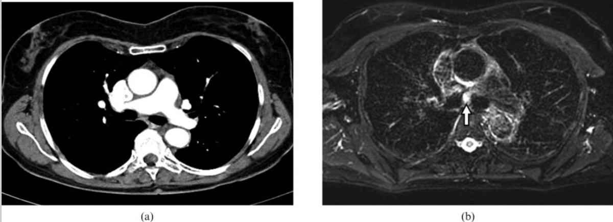
For patients with a known lung tumour, MRI excels at clarifying whether cancer has invaded the chest wall, mediastinum, or major vessels, such as the aorta, pulmonary artery, or superior vena cava5,6. High-resolution soft-tissue imaging is crucial for surgical planning, enabling surgeons to determine whether less invasive surgery is feasible or if open surgery should be avoided.
High-Risk Patient Backgrounds
Lung MRI is especially valuable in patients at higher long-term cancer risk, such as heavy smokers in screening programs, those with a history of chest irradiation, people with genetic cancer syndromes (like Li-Fraumeni or EGFR mutations), or individuals with asbestos exposure1,7. In these groups, MRI’s absence of radiation provides a safer option for repeated surveillance or follow-up imaging.
Staging and Metastasis Search
For staging lung cancer and detecting distant spread, whole-body MRI can identify small pleural, vertebral, or brain metastases that may escape detection on CT or PET/CT5,6. Identifying additional metastatic sites can significantly impact the treatment plan, informing whether curative or systemic therapy is most suitable.
Unresolved or Ambiguous Chest Symptoms
When symptoms such as persistent hemoptysis (coughing up blood), unexplained chest pain, or chronic cough remain unresolved despite X-ray or low-dose CT, MRI provides additional clarification7,8. It is particularly helpful when distinguishing between inflammatory disorders and tumours, leading to more informed diagnoses.
Monitoring Treatment Response
MRI can track changes in tumour size (such as RECIST criteria), as well as more advanced markers like apparent diffusion coefficient (ADC) and perfusion after treatment with chemotherapy, immunotherapy, stereotactic radiotherapy (SBRT), ablation, or targeted drugs9. This enables more precise and early assessment of how a cancer is responding to therapy.
For Motion-Limited or Radiation-Sensitive Patients
MRI is the imaging modality of choice for patients who are pregnant, paediatric, frail, or otherwise unable to tolerate ionising radiation1,7,10. Special respiratory-gated or ultra-short echo time (UTE) MRI sequences can produce diagnostic-quality images even in these especially sensitive situations, reducing the risks associated with traditional scans.
You can use our Know Your Risk Calculator to understand your risk of cancer in just five minutes.
How to Prepare for Your Lung MRI
Here are a few tips to help you prepare for your MRI11:
- Take your usual medications and eat normally, unless instructed to fast for specific MRI protocols. Fasting is not always required however12. Avoid heavy caffeine intake and stay hydrated.
- Inform staff about any metal implants or devices, and bring safety cards for “MR-Conditional” implants, such as pacemakers or aneurysm clips.
- Remove all metal items, including jewellery, hairpins, dental plates, and transdermal patches with foil, to prevent image distortion.
- Bring your referral, prior brain images, and insurance pre-authorisation to avoid delays.
- Wear comfortable, metal-free clothing; ask about earplugs, music, or mirror goggles if you’re claustrophobic.
- If contrast is planned, fast for 4–6 hours prior to the procedure. Inform staff about any kidney issues or past reactions to contrast agents, as safer alternatives may be available.
- Arrange an escort home if you need sedation for claustrophobia.
You can read more about preparation for Ezra’s Full Body Scan here.
What Happens During the Lung Scan?
Upon arrival for your MRI, you will need to check in and complete a screening form. This will allow you to confirm the presence of implants, allergies, and whether you might need any anxiety medication.
During the scan, you will lie down on a sliding table. A dedicated surface or phased-array coil is typically placed over the limb or region of interest13. The scan typically lasts 30-45 minutes of actual “table time”, during which the technician may acquire multiple sequences (settings). If contrast is used, you might feel a cool sensation in the arm.
You’ll hear a series of loud knocking or tapping sounds as the MRI machine works. This is completely normal. The scan usually takes about 20 to 45 minutes, and you’ll be offered earplugs or headphones to make the experience more comfortable.
You’ll stay in touch with the team via a two-way intercom and a squeeze bulb, allowing you to communicate or pause the scan if needed. If contrast is required, it’s injected halfway through, possibly causing a brief cool sensation. After the final sequence, the coil is removed, and you’re free to go.
At Ezra, our Full Body Plus scan takes around 60 minutes total, with 45 minutes of table time. Earplugs or headphones are available.
MRI Safety, Risks, & Side Effects
MRI is generally considered very safe when proper screening and protocols are followed, but certain risks and side effects should be understood:
- Metal and implants: The strong 3-Tesla magnet can pull or heat older pacemakers, aneurysm clips, or metal fragments14. Most modern “MR-Conditional” devices (like cochlear implants or pain pumps) are safe after screening, but all implants must be checked before scanning15.
- Gadolinium contrast: Macrocyclic gadolinium agents (e.g., gadobutrol) have an extremely low risk of allergic reactions or nephrogenic systemic fibrosis (NSF) when kidneys are healthy16. However, gadolinium can accumulate in tissues, and rare side effects such as headaches or skin changes have been reported. Many centres now offer contrast-free alternatives for routine follow-up17. You can read more about gadolinium contrast side effects here.
- Incidental findings: These are a common finding, identified in approximately 3-10 per cent of screening studies18. Most represent early or mild interstitial lung disease, emphysema, or benign, age-related changes.
- Claustrophobia: Anxiety inside the scanner is common. Wide-bore scanners, music, mirror goggles, or a single dose of oral sedative can help alleviate symptoms. Open MRI is an option if image detail can be sacrificed.
- Zero Ionising Radiation: MRI uses magnetic fields and radio waves, not X-rays, so there is no ionising radiation exposure, making it safer for repeated scans compared to CT scans19.
- Minor Sensations: Expect loud knocking, mild table vibration, and a brief cool flush if contrast is injected. Rare side effects include headaches, fatigue, or mild skin heating.
A deeper dive into possible side effects (such as heat, headaches, and gadolinium deposition) is available in our full guide.
At Ezra, we employ a contrast-free approach using wide-bore T3 machines to deliver a comfortable scanning experience.
Terms You Might See in Your MRI Report (And What They Might Mean)
MRI reports for lung scans often include specialised terms. Here’s a guide to some common phrases and what they mean for your diagnosis:
- Speculated margin: This describes a nodule or mass with jagged, radiating lines around its edge, giving it an appearance associated with a higher likelihood of cancer20.
- Ground glass opacity (GGO): A hazy area that does not completely block underlying blood vessels or airways20. GGOs can signal early lung cancer, infection, or inflammation.
- ADC value 0.8: The apparent diffusion coefficient (ADC) measures how freely water molecules move in tissue21. A value around 0.8 x 10⁻³ mm2/s indicates a strong restriction, often suggesting a highly cellular tumour.
- Ktrans 0.24 min-1: Ktrans is a perfusion parameter that quantifies the rate at which contrast dye enters tissue. A value of 0.24 min-1 suggests moderate blood flow into the lesion, which may provide clues about tumour activity or response to therapy22,23.
- Lung-RADS 4A: Part of the Lung Imaging Reporting and Data System, this category indicates a suspicious nodule with an elevated risk of cancer. Early follow-up, possibly including additional imaging or biopsy, is recommended24.
- Volume-doubling time: This is a calculated estimate indicating how quickly a lung nodule is growing. A shorter doubling time points toward a more aggressive lesion25.
- Pleural effusion: Pleural effusion is the abnormal accumulation of fluid between the layers of the pleura. The presence of pleural effusion can indicate inflammation, infection, or cancer spreading to the pleura20.
- Major vessel invasion: This signals that a tumour is abutting or wrapping around more than half (over 180 degrees) of a main blood vessel like the aorta or pulmonary artery. When present, it can greatly affect whether surgical removal is possible.
- Computed tomography dose index: Represents the radiation dose delivered during an associated low-dose CT scan26.
Ezra provides a radiologist-reviewed report in a non-technical and easy-to-understand format on your dashboard.
After the MRI Scan
After the MRI scan, you will be free to go home and continue with your day without any precautions27. If you received a sedative, you will need another person to pick you up. You will also not be able to drive, consume alcohol or operate heavy machinery 24 hours after the sedative.
A team of experts will review your results and determine whether a follow-up is necessary and recommend the appropriate treatment if needed. If abnormalities are found, you may undergo ongoing monitoring every 2-3 months to track recurrence. You can receive support in the form of counselling and advice on how to handle aspects like claustrophobia.
If you have a scan with us here at Ezra, you will receive your report within five to seven days and have the option to discuss it with a medical practitioner. You can also access your scan images through the online portal.
What MRI Can Show About Lung Cancer
Lesion Size and Margins: The Cornerstone of T-Staging
MRI provides precise measurements of lung tumour size and shape, distinguishing between smooth margins (often benign or indolent) and spiculated, jagged margins, which are closely linked to a higher risk of malignancy and a worse prognosis28,29. Spiculated or irregular margins also carry independent prognostic information, aiding both accurate staging and risk prediction.
Relationship to Vessels, Bronchi, and Pleura: Defining Surgical Options
A key strength of MRI lies in its ability to reveal how the tumour relates to nearby structures such as major blood vessels, airways, and the pleura6,30. Clear imaging of tumour extension or invasion determines whether the lesion can be safely removed by surgery or if vital structures are involved, making surgery unfeasible.
Cavitation or Necrosis: Clues to Tumour Type and Biopsy Planning
MRI can detect central areas of necrosis (dead tissue) or cavitation (air-filled spaces within a tumour), findings that are particularly common in squamous cell lung cancers31,32. The presence of cavitation may change biopsy plans and influence treatment by indicating aggressive biology or past therapy effects, especially in patients receiving anti-angiogenic drugs.
Diffusion Restriction and ADC Value: Markers of Tumour Cellularity and Aggression
Diffusion-weighted MRI allows for quantification of how easily water molecules move within tumour tissue, summarised as the ADC. Low ADC values (typically less than 1.0 x 10⁻³ mm²/s) signal high cellular density, an indicator of aggressive, poorly differentiated cancers, and are associated with worse histologic grade and higher tumour aggressiveness33,34.
Dynamic Contrast Curves: Characterising Tumour Vascularity
With dynamic contrast-enhanced MRI, tumours can be evaluated for how quickly and how much they “light up” after contrast injection. Malignant tumours often demonstrate rapid early enhancement with a plateau or quick washout, while a slower, progressive pattern is more characteristic of scar tissue or inflammation, helping to differentiate malignant from benign lesions35,36.
Perfusion Heterogeneity
Advanced MRI techniques can assess the variability in blood flow (perfusion heterogeneity) throughout the tumour. Regions with poor perfusion often correspond to hypoxic areas, subsets of tumours that may be more resistant to chemotherapy and radiotherapy, providing important information for individualising treatment37,38.
Ezra utilises DWI as part of our whole-body MRI scans and artificial intelligence (AI) to enhance MRI images and convert radiology reports into layman's term translations.
Types of Lung Tumours and How They Look on MRI
Adenocarcinoma (Solid/Glandular): Lung adenocarcinomas can present as small pulmonary nodules with high tissue-contrast resolution on MRI. Tumour components exhibit variable signal and enhancement patterns: aggregated tumour cells often display marked early enhancement, while fibrotic components are typically hypointense, and necrotic areas are hyperintense on T2-weighted images39.
Squamous-Cell Carcinoma: Squamous-cell carcinoma tends to occur centrally in the lungs and often manifests as a mass of central cavitation40. On MRI and perfusion studies, it usually shows strong enhancement and intense signal on DWI.
Small-Cell Lung Cancer: Small-cell lung cancer (SCLC) typically presents as a bulky hilar or mediastinal mass. On MRI, SCLC shows very low ADC values, which are often associated with extensive nodal spread at the time of diagnosis41.
Carcinoid Tumour: These tumours are generally well-defined, lobulated nodules that show strong, even contrast enhancement on MRI42. These tumours are highly vascular but tend not to invade adjacent structures, and their smooth borders help differentiate them from more aggressive cancers.
Mucinous Tumours: Mucinous lung tumours (invasive mucinous adenocarcinomas) characteristically display high signal on T2-weighted MRI due to their mucin-rich content43,44. They may also exhibit raised proton-density fat-fraction (PDFF) on specialised MRI sequences, and frequently show ground-glass or consolidative opacities with air bronchogram.
Ezra screens for over 500 conditions, including lung abnormalities.
Types of MRI Scans Used in Lung Cancer Detection
There are multiple types of MRI scans, all using different methods to give a better visualisation of lung tumours.
- T1-weighted 3D GRE sequences are fundamental for evaluating thoracic anatomy and provide sharp, high-resolution images of the lung and mediastinum45. These scans excel in post-contrast imaging, helping to delineate tumours from adjacent structures and assess subtle invasion into the chest wall, vessels, or pericardium.
- T2-weighted fast spin-echo MRI is particularly valuable for highlighting areas of increased water content, such as oedema, fluid collections, or necrosis within tumours2,5,46. These sequences are sensitive for identifying tumour-associated changes, such as central necrosis, which is especially important in squamous cell carcinoma, and can also distinguish fluid-filled spaces from solid components.
- DWI/ADC measures the motion of water molecules within tissues and is highly sensitive for detecting tumour cellularity2,5,46. Lesions with restricted diffusion typically correspond to higher cellular density, a feature of many aggressive cancers, while ADC values provide a quantitative biomarker for monitoring early response to therapies like chemotherapy and radiotherapy.
- Dynamic contrast-enhanced perfusion MRI involves rapid, time-resolved scanning after contrast injection to assess tumour vascularity22,47,48. Quantitative parameters such as Ktrans and Kep reflect how quickly and efficiently the tumour takes up and releases contrast, which can help differentiate benign from malignant lesions and predict treatment response.
- Ultra-short echo time MRI uses extremely short echo times to directly visualise lung parenchyma, overcoming the usual signal loss seen in standard MRI due to rapid signal decay in air-filled tissues49. This is especially valuable for patients who are unable to hold their breath, such as young children, frail individuals, or those with significant respiratory compromise, and can improve the detection of small nodules or subtle interstitial changes.
- Respiratory-gated or navigator-triggered sequences allow imaging during normal breathing, minimising motion artefacts from the chest wall or diaphragm45,50. This approach is well-suited for children or patients with debilitation and can be combined with other MRI sequences to deliver diagnostic-quality images without exposure to ionising radiation.
- Whole-body MRI incorporating DWI enables a comprehensive, single-session assessment for metastatic spread, including the brain, bone, and liver51. This approach can match the diagnostic accuracy of multi-modality pathways for staging and can be particularly helpful when PET/CT is contraindicated or unavailable.
Ezra uses whole-body DWI imaging to get a full picture of the body and catch any potential abnormalities.
MRI vs. Other Imaging Tests for Lung Cancer Detection
MRI Scan Cost
Ezra’s MRI Scan with Spine costs £2,395 and is currently available at their partner clinic in Marylebone, London and in Sidcup, with more locations planned in the future. You can also add a lung CT for £249 to look for lung cancer, pulmonary nodules, emphysema, and more. No referral is required, so you can book your scan directly without consulting a GP or specialist first. Most people pay out of pocket, as insurance typically does not cover self-referred scans, but you may be able to seek reimbursement depending on your policy.
Frequently Asked Questions
Does your whole body go in for a lung MRI?
Typically, only the chest needs to be imaged for a lung MRI; however, if a whole-body disease assessment or metastasis survey is required, your entire body may be scanned.
Can lung cancer be detected by MRI?
Yes, MRI can detect lung cancer and is especially useful for characterising tumours, assessing their relationships to surrounding structures, and staging, though low-dose CT remains the gold standard for initial detection.
Is MRI better than CT for lung cancer?
CT is generally superior to MRI for detecting lung cancer and nodules due to its higher sensitivity and specificity; however, MRI offers advantages for evaluating soft tissue and in certain clinical scenarios.
Can MRI show lung nodules?
MRI can reliably detect lung nodules larger than 4–6 mm and performs almost as well as CT for nodules 8–10 mm or larger; however, it is less sensitive for smaller nodules.
Why Does Ezra use MRI & CT?
Ezra uses both MRI and CT because MRI provides comprehensive, radiation-free imaging of multiple organs, while low-dose CT remains best for lung cancer screening and evaluating certain conditions that MRI cannot assess as well.
Key Takeaways
- Combine Low-Dose CT (LD-CT) with Targeted MRI: Utilising both low-dose CT and focused MRI enables the early detection of lung cancer, significantly increasing the chances of a cure when the disease is still in its early stages.
- MRI Provides Unique Functional Information Without Extra Radiation: MRI delivers critical insights into tumour biology, such as tissue diffusion and blood perfusion, without subjecting patients to additional radiation. This is particularly valuable for children, younger adults, and individuals who are sensitive to radiation or require frequent scans.
- Track Change, Not Just Size: Advances like AI-calculated volume-doubling time and Lung-RADS scoring provide more meaningful assessments of a nodule's aggressiveness than measuring diameter alone. Monitoring these dynamic changes helps identify fast-growing cancers that may require urgent intervention.
- Quality Scans Require Good Breath-Holds and Motion Control: Ensuring patients can hold their breath and remain still during MRI scans improves image sharpness, reduces the need for repeat scans, and enhances diagnostic accuracy.
- Early, Multimodal Imaging and Customised Follow-Up Improve Outcomes: A strategy that integrates multiple imaging techniques (such as LD-CT, MRI, and AI-based tools) and tailors follow-up intervals to each patient’s risk profile is shown to improve survival by detecting cancer early and closely monitoring treatment response.


References
1. Biederer J, Mirsadraee S, Beer M, et al. MRI of the lung (3/3)—current applications and future perspectives. Insights Imaging. 2012;3(4):373-386. doi:10.1007/s13244-011-0142-z
2. Usuda K, Iwai S, Funasaki A, et al. Diffusion-weighted magnetic resonance imaging is useful for the response evaluation of chemotherapy and/or radiotherapy to recurrent lesions of lung cancer. Transl Oncol. 2019;12(5):699-704. doi:10.1016/j.tranon.2019.02.005
3. Binczyk F, Prazuch W, Bozek P, et al. Radiomics and artificial intelligence in lung cancer screening. Transl Lung Cancer Res. 2021;10(2):1186-1199. doi:10.21037/tlcr-20-708
4. Biederer J, Hintze C, Fabel M. MRI of pulmonary nodules: technique and diagnostic value. Cancer Imaging. 2008;8(1):125-130. doi:10.1102/1470-7330.2008.0018
5. Hochhegger B, Marchiori E, Sedlaczek O, et al. MRI in lung cancer: a pictorial essay. Br J Radiol. 2011;84(1003):661-668. doi:10.1259/bjr/24661484
6. Khalil A, Bouhela T, Carette M. Contribution of MRI in lung cancer staging. Journal of the Belgian Society of Radiology. 2013;96(3):132. doi:10.5334/jbr-btr.234
7. MRI of the lungs and bronchi - what the study shows, its indications and benefits. August 24, 2023. Accessed July 17, 2025. https://medconsonline.com/en/blog/lung-and-bronchi-mri
8. Kauczor HU, Ley-Zaporozhan J, Ley S. Imaging of Pulmonary Pathologies. Proc Am Thorac Soc. 2009;6(5):458-463. doi:10.1513/pats.200901-002AW
9. Bainbridge H, Salem A, Tijssen RHN, et al. Magnetic resonance imaging in precision radiation therapy for lung cancer. Transl Lung Cancer Res. 2017;6(6):689-707. doi:10.21037/tlcr.2017.09.02
10. Cobben DCP, de Boer HCJ, Tijssen RH, et al. Emerging Role of MRI for Radiation Treatment Planning in Lung Cancer. Technol Cancer Res Treat. 2016;15(6):NP47-NP60. doi:10.1177/1533034615615249
11. Radiology (ACR) RS of NA (RSNA) and AC of. Magnetic Resonance Imaging (MRI) - Head. Radiologyinfo.org. Accessed July 3, 2025. https://www.radiologyinfo.org/en/info/mri-brain
12. Williams JM, Hilmes MA, Archer B, et al. Repeatability and Reproducibility of Pancreas Volume Measurements Using MRI. Sci Rep. 2020;10:4767. doi:10.1038/s41598-020-61759-9
13. Gruber B, Froeling M, Leiner T, et al. RF coils: A practical guide for nonphysicists. J Magn Reson Imaging. 2018;48(3):590-604. doi:10.1002/jmri.26187
14. Gill A, Shellock FG. Assessment of MRI issues at 3-Tesla for metallic surgical implants: findings applied to 61 additional skin closure staples and vessel ligation clips. J Cardiovasc Magn Reson. 2012;14(1):3. doi:10.1186/1532-429X-14-3
15. Potential Hazards and Risks. UCSF Radiology. January 20, 2016. Accessed March 14, 2025. https://radiology.ucsf.edu/patient-care/patient-safety/mri/potential-hazards-risks
16. Costello JR, Kalb B, Martin DR. Incidence and Risk Factors for Gadolinium-Based Contrast Agent Immediate Reactions. Top Magn Reson Imaging. 2016;25(6):257-263. doi:10.1097/RMR.0000000000000109
17. McDonald RJ, McDonald JS, Kallmes DF, et al. Gadolinium Deposition in Human Brain Tissues after Contrast-enhanced MR Imaging in Adult Patients without Intracranial Abnormalities. Radiology. 2017;285(2):546-554. doi:10.1148/radiol.2017161595
18. O’Dowd EL, Tietzova I, Bartlett E, et al. ERS/ESTS/ESTRO/ESR/ESTI/EFOMP statement on management of incidental findings from low dose CT screening for lung cancer. European Journal of Cardio-Thoracic Surgery. 2023;64(4):ezad302. doi:10.1093/ejcts/ezad302
19. Mall MA, Stahl M, Graeber SY, et al. Early detection and sensitive monitoring of CF lung disease: Prospects of improved and safer imaging. Pediatr Pulmonol. 2016;51(S44):S49-S60. doi:10.1002/ppul.23537
20. Bankier AA, MacMahon H, Colby T, et al. Fleischner Society: Glossary of Terms for Thoracic Imaging. Radiology. 2024;310(2):e232558. doi:10.1148/radiol.232558
21. Aslan A, İnan İ, Aktan A, et al. The utility of ADC measurement techniques for differentiation of low- and high-grade clear cell RCC. Pol J Radiol. 2018;83:e446-e451. doi:10.5114/pjr.2018.80207
22. Wang D, Liu S, Fu J, et al. Correlation of Ktrans derived from dynamic contrast-enhanced MRI with treatment response and survival in locally advanced NSCLC patients undergoing induction immunochemotherapy and concurrent chemoradiotherapy. J Immunother Cancer. 2024;12(6):e008574. doi:10.1136/jitc-2023-008574
23. Chen L, Zeng X, Wu Y, et al. A Study of the Correlation of Perfusion Parameters in High-Resolution GRASP MRI With Microvascular Density in Lung Cancer. J Magn Reson Imaging. 2019;49(4):1186-1194. doi:10.1002/jmri.26340
24. Morgan MA. Lung Imaging Reporting and Data System (Lung-RADS) | Radiology Reference Article | Radiopaedia.org. Radiopaedia. doi:10.53347/rID-32681
25. Jiang B, Han D, van der Aalst CM, et al. Lung cancer volume doubling time by computed tomography: A systematic review and meta-analysis. European Journal of Cancer. 2024;212:114339. doi:10.1016/j.ejca.2024.114339
26. Kalra MK, Karout L, Kiipper F de M, et al. Decoding dose descriptors for computed tomography. Radiol Bras. 57:e20230116. doi:10.1590/0100-3984.2023.0116
27. MRI scan. NHS inform. Accessed July 3, 2025. https://www.nhsinform.scot/tests-and-treatments/scans-and-x-rays/mri-scan/
28. Lee G, Park H, Lee HY, et al. Tumor Margin Contains Prognostic Information: Radiomic Margin Characteristics Analysis in Lung Adenocarcinoma Patients. Cancers (Basel). 2021;13(7):1676. doi:10.3390/cancers13071676
29. Yi CA, Jeon TY, Lee KS, et al. 3-T MRI: Usefulness for Evaluating Primary Lung Cancer and Small Nodules in Lobes Not Containing Primary Tumors. American Journal of Roentgenology. 2007;189(2):386-392. doi:10.2214/AJR.07.2082
30. Purandare NC, Rangarajan V. Imaging of lung cancer: Implications on staging and management. Indian J Radiol Imaging. 2015;25(2):109-120. doi:10.4103/0971-3026.155831
31. Calvetti L, Aprile G. Influence and mechanism of lung cavitation development on antiangiogenic therapy: is cavitation the new caveat? Transl Lung Cancer Res. 2019;8(4):319-322. doi:10.21037/tlcr.2019.08.19
32. Nishino M, Cryer SK, Okajima Y, et al. Tumoral cavitation in patients with non-small-cell lung cancer treated with antiangiogenic therapy using bevacizumab. Cancer Imaging. 2012;12(1):225-235. doi:10.1102/1470-7330.2012.0027
33. Liu H, Liu Y, Yu T, et al. Evaluation of apparent diffusion coefficient associated with pathological grade of lung carcinoma, before therapy. Journal of Magnetic Resonance Imaging. 2015;42(3):595-601. doi:10.1002/jmri.24823
34. Zhang F, Zhou Z, Tang D, et al. Diffusion-weighted MRI in solitary pulmonary lesions: associations between apparent diffusion coefficient and multiple histopathological parameters. Sci Rep. 2018;8(1):11248. doi:10.1038/s41598-018-29534-z
35. Donmez FY, Yekeler E, Saeidi V, et al. Dynamic Contrast Enhancement Patterns of Solitary Pulmonary Nodules on 3D Gradient-Recalled Echo MRI. American Journal of Roentgenology. 2007;189(6):1380-1386. doi:10.2214/AJR.07.2429
36. Yoo MR, Whang SH, Park CH, et al. Dynamic Contrast-Enhanced CT in Advanced Lung Cancer after Chemotherapy with/without Radiation Therapy: Can It Predict Treatment Responsiveness of the Tumor? Journal of the Korean Society of Radiology. 2013;69(2):131-138. doi:10.3348/jksr.2013.69.2.131
37. Tao X, Wang L, Hui Z, et al. DCE-MRI Perfusion and Permeability Parameters as predictors of tumor response to CCRT in Patients with locally advanced NSCLC. Sci Rep. 2016;6(1):35569. doi:10.1038/srep35569
38. Bashir U, Siddique MM, Mclean E, et al. Imaging Heterogeneity in Lung Cancer: Techniques, Applications, and Challenges. American Journal of Roentgenology. 2016;207(3):534-543. doi:10.2214/AJR.15.15864
39. Awaya H, Matsumoto T, Honjo K, et al. A preliminary study of discrimination among the components of small pulmonary nodules by MR imaging: correlation between MR images and histologic appearance. Radiat Med. 2000;18(1):29-38.
40. Yang M, Shi L, Huang T, et al. Value of contrast-enhanced magnetic resonance imaging-T2WI-based radiomic features in distinguishing lung adenocarcinoma from lung squamous cell carcinoma with solid components >8 mm. Journal of Thoracic Disease. 2023;15(2). doi:10.21037/jtd-23-142
41. Christensen TN, Langer SW, Villumsen KE, et al. 18F-fluorothymidine (FLT)-PET and diffusion-weighted MRI for early response evaluation in patients with small cell lung cancer: a pilot study. Eur J Hybrid Imaging. 2020;4:2. doi:10.1186/s41824-019-0071-5
42. Megahed M, Sharif R, Abdeen Y. Synchronous bilateral typical pulmonary carcinoid tumours diagnosed by robotic navigation bronchoscopy: A unique case. Respirol Case Rep. 2024;12(11):e70055. doi:10.1002/rcr2.70055
43. Pan X, Fang R, Zhang B, et al. Pathological and imaging features of pulmonary invasive mucinous adenocarcinoma—a retrospective cohort study. Transl Lung Cancer Res. 2024;13(6):1376-1382. doi:10.21037/tlcr-24-526
44. Ogusu S, Takahashi K, Hirakawa H, et al. Primary Pulmonary Colloid Adenocarcinoma: How Can We Obtain a Precise Diagnosis? Intern Med. 2018;57(24):3637-3641. doi:10.2169/internalmedicine.1153-18
45. Wang YXJ, Lo GG, Yuan J, et al. Magnetic resonance imaging for lung cancer screen. J Thorac Dis. 2014;6(9):1340-1348. doi:10.3978/j.issn.2072-1439.2014.08.43
46. Broncano J, Steinbrecher K, Marquis KM, et al. Diffusion-weighted Imaging of the Chest: A Primer for Radiologists. RadioGraphics. 2023;43(7):e220138. doi:10.1148/rg.220138
47. Hatzoglou V, Tisnado J, Mehta A, et al. Dynamic contrast‐enhanced MRI perfusion for differentiating between melanoma and lung cancer brain metastases. Cancer Med. 2017;6(4):761-767. doi:10.1002/cam4.1046
48. Guo W, Lv B, Yang T, et al. Role of Dynamic Contrast-Enhanced Magnetic Resonance Imaging Parameters and Extracellular Volume Fraction as Predictors of Lung Cancer Subtypes and Lymph Node Status in Non-Small-Cell Lung Cancer Patients. J Cancer. 2023;14(16):3108-3116. doi:10.7150/jca.88367
49. Takahashi M, Togao O, Obara M, et al. Ultra-short echo time (UTE) MR imaging of the lung: Comparison between normal and emphysematous lungs in mutant mice. J Magn Reson Imaging. 2010;32(2):326-333. doi:10.1002/jmri.22267
50. Renz DM, Herrmann KH, Kraemer M, et al. Ultrashort echo time MRI of the lung in children and adolescents: comparison with non-enhanced computed tomography and standard post-contrast T1w MRI sequences. Eur Radiol. 2022;32(3):1833-1842. doi:10.1007/s00330-021-08236-7
51. nihr_wp. Whole-body MRI scans are as accurate as standard imaging pathways for lung cancer staging. NIHR Evidence. doi:10.3310/signal-000796