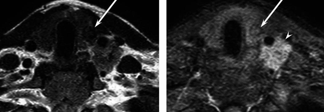
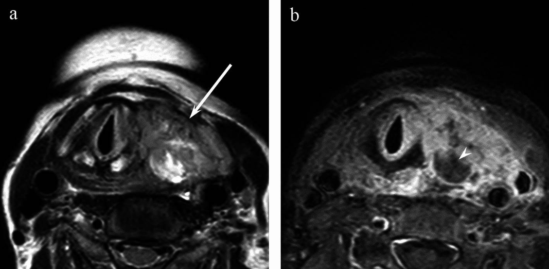
A thyroid MRI (magnetic resonance imaging) is a non-invasive imaging test that provides high-resolution images of the thyroid gland and adjacent neck structures, making it a valuable tool in modern thyroid disease assessment. This procedure is particularly helpful in staging known or suspected thyroid malignancy, monitoring for recurrence, and evaluating complex cases such as large goitres, cysts, or nodules when other imaging modalities such as ultrasound or computed tomography (CT) are insufficient1.
A thyroid MRI might be recommended for several clinical reasons:
Here are a few tips to help you prepare for your MRI5:
You can read more about preparation for Ezra’s MRI Scan with Spine here.
Upon arrival for your MRI, you will need to check in and complete a screening form. This will allow you to confirm the presence of implants, allergies, and whether you might need any anxiety medication.
During the scan, you will lie down on a sliding table. A dedicated surface or phased-array coil is typically placed over the limb or region of interest6. Your head will be nestled in a small cushion that will keep you still. The scan typically lasts 30-45 minutes of actual “table time”, during which the technician may acquire multiple sequences (settings). Expect loud knocking noises (up to 110 dB); earplugs or headphones are provided to reduce discomfort. It’s normal to feel mild table vibrations.
You’ll stay in touch with the team via a two-way intercom and a squeeze bulb, allowing you to communicate or pause the scan if needed. If contrast is required, it’s injected halfway through, possibly causing a brief cool sensation. After the final sequence, the coil is removed, and you’re free to go.
At Ezra, our MRI Scan with Spine scan takes around 60 minutes total, with 45 minutes of table time. Earplugs or headphones are available.
After the scan, you will be contacted by a medical provider working with Ezra within roughly a week. On the day of the appointment, you will receive a copy of your report and access to your scanned images through the online portal.
MRI is generally considered very safe when proper screening and protocols are followed, but certain risks and side effects should be understood:
A deeper dive into possible side effects (such as heat, headaches, and gadolinium deposition) is available in our full guide.
At Ezra, we employ a contrast-free approach using wide-bore T3 machines to deliver a comfortable scanning experience.
MRI reports of the thyroid include specific terms that help clinicians assess the nature of a lesion or condition. Some common terms (and their meanings) include:
Heterogeneous Signal: Indicates the nodule or thyroid tissue has mixed signal intensity, often due to the presence of different tissue types. This can raise suspicion for malignancy, but is nonspecific13.
Extrathyroidal Extension (ETE): Refers to the spread of a tumour beyond the thyroid gland itself into surrounding tissues, which is typically a sign of more aggressive disease.
Apparent Diffusion Coefficient: A numerical value from diffusion-weighted imaging (DWI); low ADC values are often associated with cancer because malignant tissue tends to restrict water movement.
Restricted Diffusion: Seen when water molecule movement is limited within a lesion, suggesting high cellularity, which is suspicious for malignant nodules.
Enhancement: Refers to how a thyroid lesion absorbs contrast dye. Malignant tissue often enhances more than benign tissue, suggesting increased vascularity or abnormal tissue behaviour.
Mass Effect: Indicates that a thyroid lesion is exerting pressure on or displacing adjacent structures, which may signify substantial size or aggressive growth.
Ezra provides a radiologist-reviewed report in a non-technical and easy-to-understand format on your dashboard.
After the MRI scan, you will be free to go home and continue with your day without any precautions14. If you received a sedative, you will need another person to pick you up. You will also not be able to drive, consume alcohol, or operate heavy machinery 24 hours after the sedative.
A team of experts will review your results and determine whether a follow-up is necessary and recommend the appropriate treatment if needed. If abnormalities are found, you may undergo ongoing monitoring every 2-3 months to track recurrence. You can receive support in the form of counselling and advice on how to handle aspects like claustrophobia.
If you have a scan with us here at Ezra, you will receive your report within five to seven days and have the option to discuss it with a medical practitioner. You can also access your scan images through the online portal.
MRI is a valuable tool in thyroid imaging, offering detailed insights into nodule composition, tumour spread, invasion of surrounding tissues, and lymph node involvement. It can detect imaging characteristics that help differentiate malignant from benign thyroid nodules.
MRI can show whether a thyroid nodule is solid, cystic, or mixed15. This distinction is foundational to risk assessment, as solid elements raise concern for cancer, while purely cystic nodules are almost always benign.
MRI accurately detects extrathyroidal extension, which means the tumour has spread beyond the thyroid gland itself into surrounding tissues like muscle or fat1,15. This finding signifies a more advanced or aggressive disease and affects treatment plans significantly.
MRI can clarify whether cancer is invading key structures near the thyroid, such as the windpipe, nerves, and major blood vessels, which is critical for surgical planning and prognosis1,16.
MRI is sensitive in detecting suspicious lymph nodes in the neck1,15,16. Enlarged or abnormal appearing nodes may indicate metastatic spread, necessitating further evaluation or biopsy.
Tissue features like restricted diffusion and pronounced contrast enhancement often suggest malignancy in a thyroid lesion15. Malignant tumours typically show lower ADC values and greater enhancement compared to benign nodules.
MRI can distinguish between different types of thyroid tumours by revealing their structural and signal characteristics. Each tumour type displays specific features helpful for diagnosis and management decisions.
PTC is the most common thyroid cancer. On MRI, it usually appears as a solid mass with irregular margins, showing restricted diffusion (low ADC values) and is sometimes associated with lymph node metastasis. The solid components often display a hypointense signal on T2-weighted images compared to benign nodules. Ill-defined margins and extrathyroidal extension are also frequent.

Follicular carcinomas often appear brighter than normal thyroid tissue on T2-weighted scans and tend to stand out after contrast is given. The shape may be irregular, with edges that are not clearly defined. Sometimes you may see a thin dark rim around the tumour, but if this rim is incomplete or broken, it suggests the tumour has pushed through its capsule and may also show areas where it is invading blood vessels18,19. These changes help doctors distinguish follicular carcinoma from less harmful lumps, even when their signal is otherwise similar.
The solid components of anaplastic thyroid carcinoma frequently show hyperintensity on T2-weighted images compared to the spinal cord17. It also often presents with ill-defined margins and shows a higher incidence of extension out of the thyroid, tracheal, and oesophageal invasion, vascular invasion, and venous thrombosis, reflecting their aggressive nature.

Benign thyroid nodules are usually well-defined, homogenous lesions that do not enhance (or only mildly enhance) after contrast administration15. Simple cystic nodules have very high T2 signal intensity and lack solid components or invasive features. Solid benign nodules show higher T2 signal and ASC values than malignancies, reflecting their lower cellularity.
Ezra screens for over 500 conditions, including the brain.
There are several types of MRI scans, each providing unique information to help visualise the thyroid gland and detect cancer3,15.
Ezra’s MRI Scan with Spine costs £2,395 and is currently available at their partner clinic in Marylebone, London and in Sidcup, with more locations planned in the future. No referral is required, so you can book your scan directly without consulting a GP or specialist first. Most people pay out-of-pocket, as insurance typically does not cover self-referred scans, but you may be able to seek reimbursement depending on your policy.
Ultrasound is usually the first and most cost-effective test for thyroid cancer, but MRI can provide extra detail for larger or complex cases, especially involving invasion outside the thyroid.
A biopsy is still needed to definitively diagnose thyroid cancer, even if MRI findings are suspicious.
MRI does not use radiation and is considered safe for repeated use.


1. Hoang JK, Branstetter BF, Gafton AR, Lee WK, Glastonbury CM. Imaging of thyroid carcinoma with CT and MRI: approaches to common scenarios. Cancer Imaging. 2013;13(1):128-139. doi:10.1102/1470-7330.2013.0013
2. Bomeli SR, LeBeau SO, Ferris RL. Evaluation of a thyroid nodule. Otolaryngol Clin North Am. 2010;43(2):229-238. doi:10.1016/j.otc.2010.01.002
3. King AD. Imaging for staging and management of thyroid cancer. Cancer Imaging. 2008;8(1):57-69. doi:10.1102/1470-7330.2008.0007
4. Park JO, Kim JH, Joo YH, et al. Guideline for the Surgical Management of Locally Invasive Differentiated Thyroid Cancer From the Korean Society of Head and Neck Surgery. Clin Exp Otorhinolaryngol. 2023;16(1):1-19. doi:10.21053/ceo.2022.01732
5. Radiology (ACR) RS of NA (RSNA) and AC of. Magnetic Resonance Imaging (MRI) - Head. Radiologyinfo.org. Accessed July 3, 2025. https://www.radiologyinfo.org/en/info/mri-brain
6. Gruber B, Froeling M, Leiner T, Klomp DWJ. RF coils: A practical guide for nonphysicists. J Magn Reson Imaging. 2018;48(3):590-604. doi:10.1002/jmri.26187
7. Gill A, Shellock FG. Assessment of MRI issues at 3-Tesla for metallic surgical implants: findings applied to 61 additional skin closure staples and vessel ligation clips. J Cardiovasc Magn Reson. 2012;14(1):3. doi:10.1186/1532-429X-14-3
8. Potential Hazards and Risks. UCSF Radiology. January 20, 2016. Accessed March 14, 2025. https://radiology.ucsf.edu/patient-care/patient-safety/mri/potential-hazards-risks
9. Costello JR, Kalb B, Martin DR. Incidence and Risk Factors for Gadolinium-Based Contrast Agent Immediate Reactions. Top Magn Reson Imaging. 2016;25(6):257-263. doi:10.1097/RMR.0000000000000109
10. McDonald RJ, McDonald JS, Kallmes DF, et al. Gadolinium Deposition in Human Brain Tissues after Contrast-enhanced MR Imaging in Adult Patients without Intracranial Abnormalities. Radiology. 2017;285(2):546-554. doi:10.1148/radiol.2017161595
11. Drake T, Gravely A, Westanmo A, Billington C. Prevalence of Thyroid Incidentalomas from 1995 to 2016: A Single-Center, Retrospective Cohort Study. J Endocr Soc. 2019;4(1):bvz027. doi:10.1210/jendso/bvz027
12. Mall MA, Stahl M, Graeber SY, Sommerburg O, Kauczor HU, Wielpütz MO. Early detection and sensitive monitoring of CF lung disease: Prospects of improved and safer imaging. Pediatr Pulmonol. 2016;51(S44):S49-S60. doi:10.1002/ppul.23537
13. Smith D. Incidental thyroid nodule | Radiology Reference Article | Radiopaedia.org. Radiopaedia. doi:10.53347/rID-69951
14. MRI scan. NHS inform. Accessed July 3, 2025. https://www.nhsinform.scot/tests-and-treatments/scans-and-x-rays/mri-scan/
15. Noda Y, Kanematsu M, Goshima S, et al. MRI of the Thyroid for Differential Diagnosis of Benign Thyroid Nodules and Papillary Carcinomas. American Journal of Roentgenology. 2015;204(3):W332-W335. doi:10.2214/AJR.14.13344
16. Diagnosis of Papillary Thyroid Cancer. Accessed October 8, 2025. https://www.thyroidcancer.com/thyroid-cancer/papillary/diagnosis
17. Maeda T, Kato H, Ando T, et al. MRI features of histological subtypes of thyroid cancer in comparison with CT findings: differentiation between anaplastic, poorly differentiated, and papillary thyroid carcinoma. Jpn J Radiol. 2025;43(2):210-218. doi:10.1007/s11604-024-01660-x
18. Guan YB, Xie BK, Yuan XP, Li HG. [Diagnosis of thyroid carcinoma by magnetic resonance imaging (MRI)]. Ai Zheng. 2003;22(7):739-744.
19. Hwang H, Park YN, Shim YS, Byun SS, Kim HS. Characteristic Dynamic Enhancement Pattern of MR Imaging for Malignant Thyroid Tumor. Preliminary Report. Neuroradiol J. 2011;24(3):392-394. doi:10.1177/197140091102400307