We find potential cancer in 6% of our members. 46% find an actionable clinical finding.
Mike Mettler, 45.
As a father with a family history of cancer, Mike has made it his mission to take control of his health. Mike's Ezra scan found cancer in his kidneys before it was a problem.
"A large part of the credit for this great prognosis goes to early detection: given that the tumor was found so early, it was easier to remove surgically, and any spread is unlikely"
Holly Hollings, 72.
Holly Hollings, two-time cancer survivor, learnt about her early-stage uterus cancer after her Ezra Scan showed an abnormal finding.
“Most people diagnosed with cancer twice can’t say cancer and lucky in the same sentence. I'm so thankful to have caught these cancers early.”
Sam Hires, 72.
Sam's Ezra Scan found Stage 3 kidney cancer - but luckily he found it in time. Thanks to his proactive approach to health Sam will continue living a happy and active life. “I’ve always taken my health seriously so I was shocked that the Ezra scan showed stage 3 kidney tumors on both kidneys. My physician says taking the scan has given me a chance for a longer life.”
Oops! Something went wrong while submitting the form.
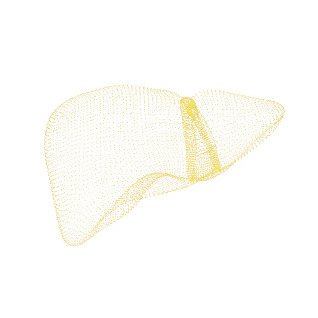
Hepatomegaly
Hepatomegaly means that the liver is enlarged. In adults, the liver normally has a span around 15 cm. The causes for hepatomegaly are varied and include infection, heart failure, cirrhosis and toxin consumption (i.e. alcohol, medication such as Tylenol, and supplements).
Abdomen
Hepatocellular adenoma
Hepatocellular adenoma is an uncommon, benign (non-cancerous) liver lesion. It has been associated with the use of estrogen-containing medications (e.g. oral contraceptives), anabolic steroids, obesity, metabolic syndrome (e.g. diabetes), and certain genetic syndromes. Hepatic adenomas may be asymptomatic (do not cause symptoms) or may cause symptoms of right upper abdominal pain, nausea, and the sensation of feeling full.
Abdomen
Hepatosplenomegaly
Hepatosplenomegaly means that both the liver and the spleen are enlarged. The liver and spleen are connected via the portal vein system. When there is a problem in one organ, it can also cause a problem in the other. The causes for hepatosplenomegaly are varied, and include obesity, infection, anemia, heart failure, cirrhosis, and leukemia/lymphoma (blood cell cancers).
Abdomen
Focal nodular hyperplasia
Focal nodular hyperplasia (FNH) is a benign (non-cancerous) lesion that consists of liver cells surrounding a central scar. While the exact cause of FNH is unknown, it is thought to occur as a response to arterial malformations (either acquired or present at birth) within the liver. FNH is the second most prevalent liver lesion (the first being hemangioma), usually found incidentally on imaging, and typically does not cause symptoms or require treatment.
Abdomen
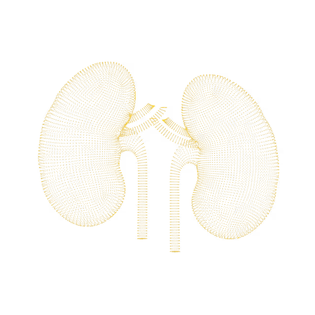
Iron overload in the liver
Iron is an important nutrient that helps the hemoglobin in blood cells carry oxygen to the body's organs and tissues. Iron overload (hemochromatosis) is a disorder in which the body has too much iron build-up. This extra iron is stored in the joints and organs; if this process is not controlled, it can cause joint and organ damage and/or failure.
Abdomen
Liver mass
Masses are growths in the liver. They could be harmless and benign or something more significant such as cancer (e.g. hepatocellular carcinoma).
Abdomen
Polycystic liver disease
Polycystic liver disease (PLD) is characterized by the presence of multiple fluid-filled liver cysts. Although a clear definition of PLD is absent, current literature defines PLD as having more than 20 liver cysts.
Abdomen
Riedel's lobe
The liver has two main portions - one right lobe and one left lobe. Sometimes the right lobe can have an anatomic variant where it appears to have a “tongue” that is still part of the right lobe and is called “Riedel's lobe.” It is a benign (non-cancerous) condition and no further evaluation or follow-up is needed.
Abdomen
Liver granuloma
A granuloma (a non-cancerous lesion that contains small abnormal clumps of cells) forms as a result of an inflammatory reaction in body tissues. Granulomas that are found in the liver are associated with inflammation caused by infection (e.g. mycobacterial infection), liver disorders (e.g. primary biliary cholangitis), and noninfectious causes (e.g. sarcoidosis, medication-induced liver injury).
Abdomen
Liver metastases
Liver metastases are lesions seen in the liver caused by the spread of cancer from other organs to the liver.
We simplify your health insights so you can quickly take action to improve your health. You can view risk levels for each region of your body and book a call with your Ezra Medical Provider for further insights into your body.
See your reports and images in MyEzra
Book a video consultation to dive deeper into your results