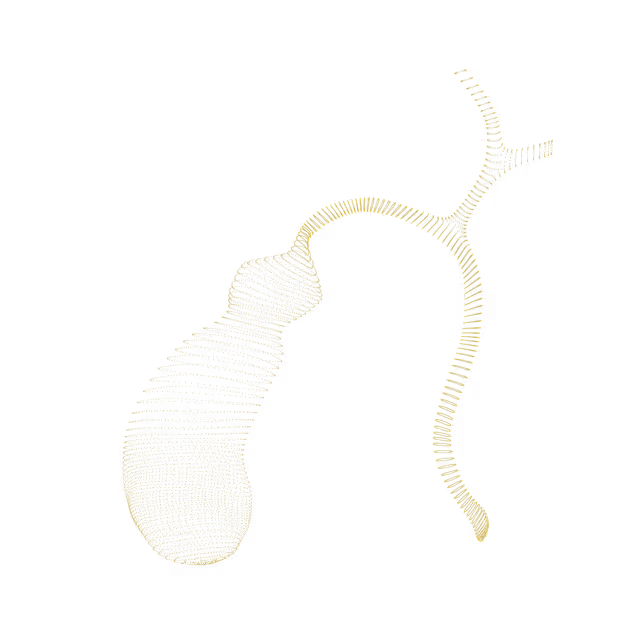
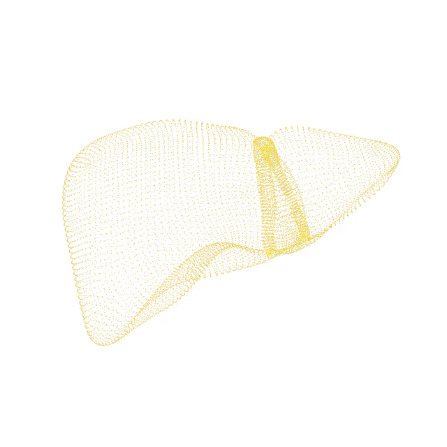
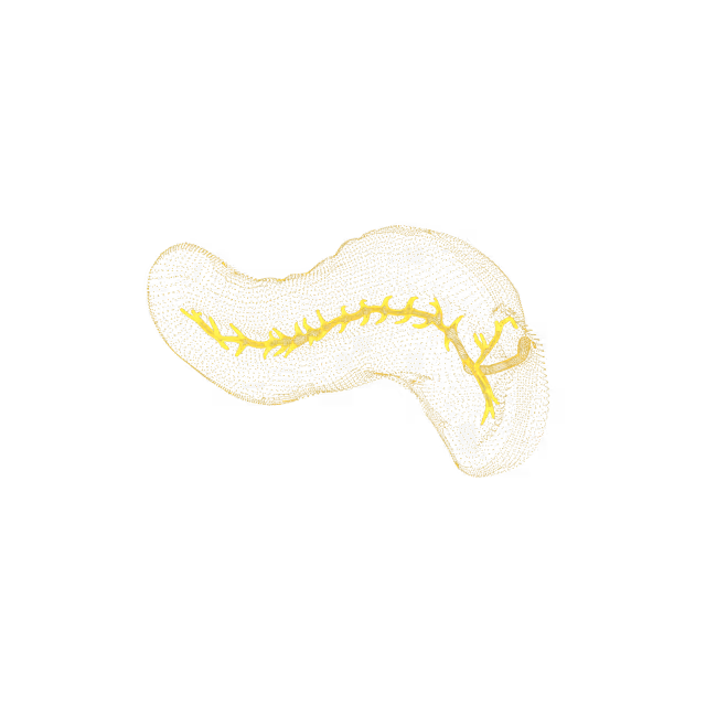
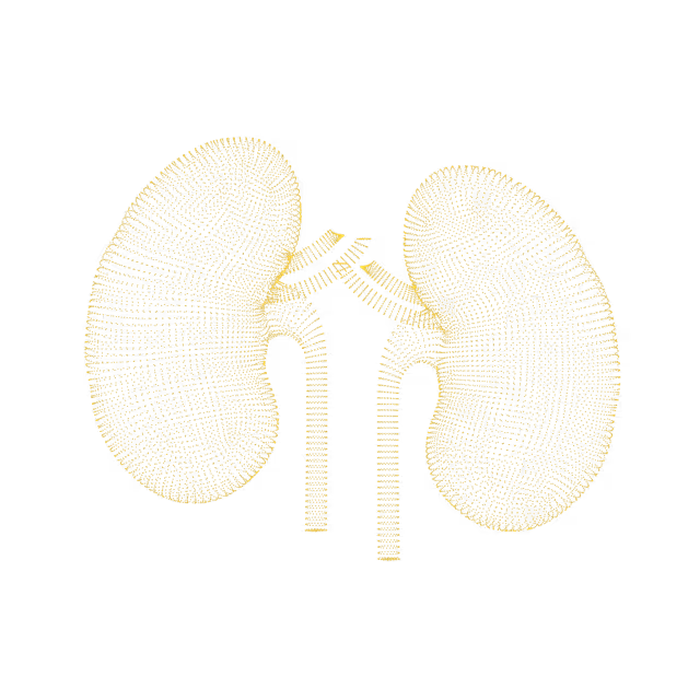
We find potential cancer in 6% of our members. 46% find an actionable clinical finding.
Mike Mettler, 45.
As a father with a family history of cancer, Mike has made it his mission to take control of his health. Mike's Ezra scan found cancer in his kidneys before it was a problem.
"A large part of the credit for this great prognosis goes to early detection: given that the tumor was found so early, it was easier to remove surgically, and any spread is unlikely"
Holly Hollings, 72.
Holly Hollings, two-time cancer survivor, learnt about her early-stage uterus cancer after her Ezra Scan showed an abnormal finding.
“Most people diagnosed with cancer twice can’t say cancer and lucky in the same sentence. I'm so thankful to have caught these cancers early.”
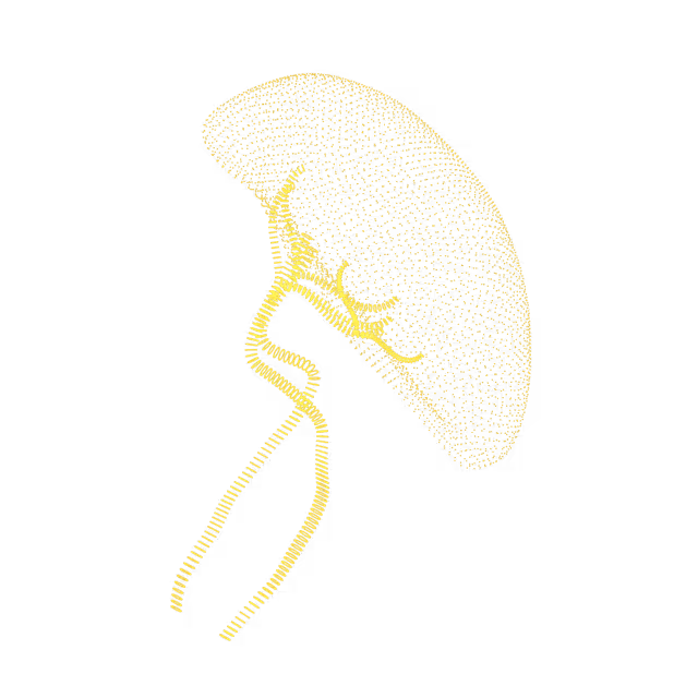
Sam Hires, 72.
Sam's Ezra Scan found Stage 3 kidney cancer - but luckily he found it in time. Thanks to his proactive approach to health Sam will continue living a happy and active life. “I’ve always taken my health seriously so I was shocked that the Ezra scan showed stage 3 kidney tumors on both kidneys. My physician says taking the scan has given me a chance for a longer life.”
Oops! Something went wrong while submitting the form.
Cervical fibroids
Cervical fibroids (also called cervical myomas, leiomyomas of the cervix) are abnormal growths of muscle tissue in the cervix. They are thought to be caused by an excess of estrogen, a hormone involved in cellular growth, and may also develop due to an imbalance between estrogen and progesterone. Cervical fibroids are benign (non-cancerous) and are usually asymptomatic (do not cause symptoms), but sometimes can be the cause of abnormal vaginal bleeding, painful sexual intercourse, and abdominal/pelvic pain.
Pelvis
Expantion of the endocervical canal
The endocervical cavity (canal) is the space connecting the vagina with the uterus. Sometimes it can be more expanded or wider than usual. It is unclear what causes this finding, but could be associated with history of vaginal birth delivery.
Pelvis
Nabothian cyst
Nabothian cysts (also called mucinous retention cysts or epithelial inclusion cysts) are mucus collections that form a sac on the cervix. They can be caused by childbirth or minor physical trauma to the cervix. Nabothian cysts are benign (non-cancerous) and are usually asymptomatic (do not cause symptoms), but sometimes can be the cause of pain or a bothersome feeling of fullness in the vagina.
Pelvis
Fluid within the cervical cavity
The cervical cavity (canal) connects the interior of the vagina with the uterus. Small amounts of fluid or mucus in the cervical cavity is a normal finding. No follow-up is indicated.
Pelvis
Bladder stones
Sometimes the salts and minerals in the urine can crystalize and form bladder stones. Factors that increase the risk of developing bladder stones include inflammation of the bladder caused by infections, foreign material present within the bladder, kidney stones, and underlying conditions (e.g. prostate gland enlargement, neurogenic bladder) that affect the bladder's ability to hold, store or eliminate urine. Most bladder stones less than or equal to 5 mm in diameter pass out of the body spontaneously and asymptomatically (with no symptoms). If a bladder stone does get stuck or irritates the bladder wall, it can cause symptoms of lower abdominal pain, difficulty urinating or interrupted urine flow, bloody urine, and painful or frequent urination.
Pelvis
Bladder diverticulum
A bladder diverticulum forms when some of the bladder lining pokes through a weak part in the bladder wall. A bladder diverticulum can either be congenital (from birth) or acquired (present later in life). Acquired bladder diverticula (when there is typically more than 1 diverticulum present) are most often caused by a blockage in the bladder outlet (such as from a swollen prostate or scars in the urethra [the tube that carries urine from the bladder out of the body]), the bladder not working well due to nerve injury or, rarely, from prior bladder surgery. Acquired diverticula are most often seen in older men, who tend to get bladder outlet blocks.
Pelvis
Bladder wall calcification
There are several possible causes of bladder wall calcification including cystitis (inflammation of the bladder), radiation to the area, schistosomiasis (a disease caused by parasitic worms that live in infested water located in tropical and subtropical regions), tuberculosis, and neoplasia (cancerous or non-cancerous growth). A diagnosis can usually be obtained from a combination of history, clinical examination, appropriate laboratory studies, and imaging of the bladder calcification and remaining urinary tract.
Pelvis
Collapsed bladder
A collapsed, or incompletely distended bladder, indicates that at the time of MRI examination, the bladder was not full of urine. This finding does not indicate any underlying condition.
Pelvis
Hydroureter
Hydroureter refers to dilation of the ureter(s), the narrow tube urine travels down from the kidneys into the bladder. It is most often caused by obstruction of urine outflow due to blockage of the ureter(s) by calculi (stones), chronic inflammation, neoplasia (cancerous or noncancerous growth), or accidental ligation during surgery.Symptoms are variable, but may include pain, either in the side and/or back (known as flank pain), nausea, and/or vomiting. Treatment is based on the cause.
Pelvis
Median lobe hypertrophy resulting in intravesical prostatic protrusion
The median lobe is located between the ejaculatory ducts and the urethra (the tube that carries urine from the bladder out of the body) in the central zone of the prostate. Hypertrophy refers to an increase in the size and number of cells in this part of the prostate. When there is overgrowth of the prostatic median lobe into the bladder, it can cause bladder outlet obstruction and related storage and voiding symptoms. While minimal hypertrophic changes may not cause symptoms, progression of these changes (also known as benign prostatic hyperplasia or BPH) may cause lower urinary tract symptoms including needing to urinate often (especially at night), difficulty starting to urinate and having a weak urine stream.
We simplify your health insights so you can quickly take action to improve your health. You can view risk levels for each region of your body and book a call with your Ezra Medical Provider for further insights into your body.
See your reports and images in MyEzra
Book a video consultation to dive deeper into your results