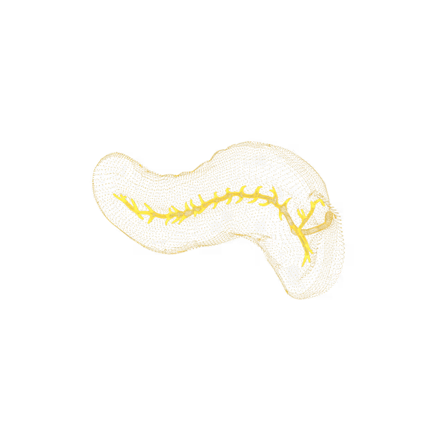
We find potential cancer in 6% of our members. 46% find an actionable clinical finding.
Mike Mettler, 45.
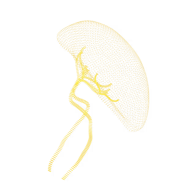
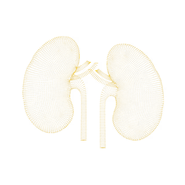
As a father with a family history of cancer, Mike has made it his mission to take control of his health. Mike's Ezra scan found cancer in his kidneys before it was a problem.
"A large part of the credit for this great prognosis goes to early detection: given that the tumor was found so early, it was easier to remove surgically, and any spread is unlikely"
Holly Hollings, 72.
Holly Hollings, two-time cancer survivor, learnt about her early-stage uterus cancer after her Ezra Scan showed an abnormal finding.
“Most people diagnosed with cancer twice can’t say cancer and lucky in the same sentence. I'm so thankful to have caught these cancers early.”
Sam Hires, 72.
Sam's Ezra Scan found Stage 3 kidney cancer - but luckily he found it in time. Thanks to his proactive approach to health Sam will continue living a happy and active life. “I’ve always taken my health seriously so I was shocked that the Ezra scan showed stage 3 kidney tumors on both kidneys. My physician says taking the scan has given me a chance for a longer life.”
Oops! Something went wrong while submitting the form.
Subchondral cyst
A subchondral cyst is a fluid-filled space inside a joint that extends from one of the bones that forms the joint. This type of bone cyst is caused by degenerative joint disease, the wear and tear on joints that develops over decades of use. The treatment depends on the degree of symptoms caused by the cyst.
Pelvis
Arthritic changes of the pelvic bone
Arthritis is the swelling and tenderness of one or more of your joints. Common symptoms include pain, stiffness and decreased range of motion. Symptoms may vary in severity and can fluctuate or progress/get worse over time. Treatments vary depending on the type of arthritis, but the main goal of treatment is to reduce symptoms and preserve joint function, mobility and quality of life.
Pelvis
Pelvic kidney
A pelvic kidney is when one or both kidneys stay in the pelvis and do not move into their proper position during fetal development. A normal variant is an atypical finding that is seen but has no clinical significance, and is considered within the spectrum of normal findings. No further follow-up or evaluation is needed for this finding.
Pelvis
Sacral chordoma
A lesion is an abnormal “spot” on the radiology images. There is a lesion noted on the coccyx (tailbone), based on your MRI images. Bone lesions may be either non-cancerous or malignant (cancerous). Sacral chordoma is a type of malignant tumor of the sacrum (the shield-shaped bone that forms the back part of the pelvis). Symptoms, if present, can include bowel, bladder, and sexual dysfuction. Pain with any sitting can also occur as the tumor grows.
Pelvis
Endometrial cyst
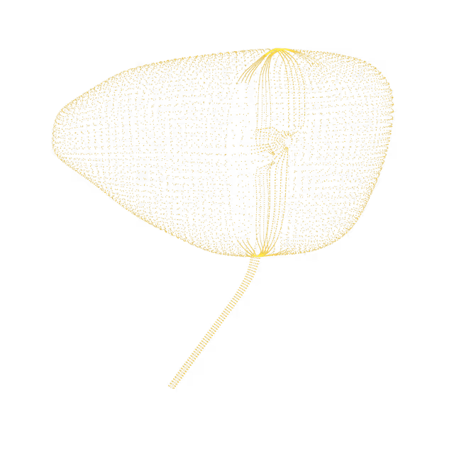
The endometrium is the inner lining of the uterus. A cyst is a sac-like pocket of membranous tissue that contains fluid, air, or other substances. Cysts can grow almost anywhere in the body. There are many different types of cysts. Most cysts are benign (non-cancerous). Whether a cyst needs treatment depends on a number of factors, including the type of cyst, the location, if the cyst is causing pain or discomfort, and whether the cyst is infected.
Pelvis
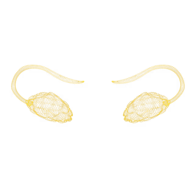
Uterus didelphys
Uterus didelphys (double uterus) is a type of uterine anomaly that is present from birth (congenital) in which a person has two uteruses. Each uterus has its own fallopian tube and ovary. Uterus didelphys is rare and only affects about 0.3% of the population. In most cases, uterine didelphys is incidentally discovered (found in passing when looking for something else) when the pelvis is imaged.
Pelvis
Calcification of the prostate
Prostate calcifications (deposits of calcium in prostate tissue) are commonly seen in men as they age. Although the exact cause is not clearly understood, experts suggest the calcifications occur as a result of chronic inflammation of the prostate. Other potential causes include diabetes, infection, benign prostatic hypertrophy (BPH), radiation therapy, previous urethral stent placement/surgery, and prostate cancer.
Pelvis
Prostate cyst
Prostatic cysts are abnormal fluid filled sacs that form in the prostate. They are a relatively common, benign finding. Cysts usually do not cause symptoms or harm the function of the prostate. Treatment is not needed for simple prostate cysts that do not cause any symptoms.
Pelvis
Prostate lesion
A prostate lesion is an area of tissue that has been damaged by injury or disease within the prostate. Prostate lesions could be benign (non-cancerous) or malignant (cancerous).
Pelvis
Stranding in prostate tissue
MRI imaging is very sensitive and it is not always clear how to interpret what is being visualized on imaging. The interpretation by the radiologist raises the possibility that there is inflammation or scarring noted in one area of your prostate, but that the cause is unclear.Inflammation and scarring in an area of the prostate could be representative of prostatitis (inflammation of the prostate). This interpretation is based solely on the MRI images, and should be taken in context with other important clinical information.
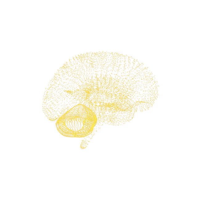
We simplify your health insights so you can quickly take action to improve your health. You can view risk levels for each region of your body and book a call with your Ezra Medical Provider for further insights into your body.
See your reports and images in MyEzra
Book a video consultation to dive deeper into your results