We find potential cancer in 6% of our members. 46% find an actionable clinical finding.
Mike Mettler, 45.
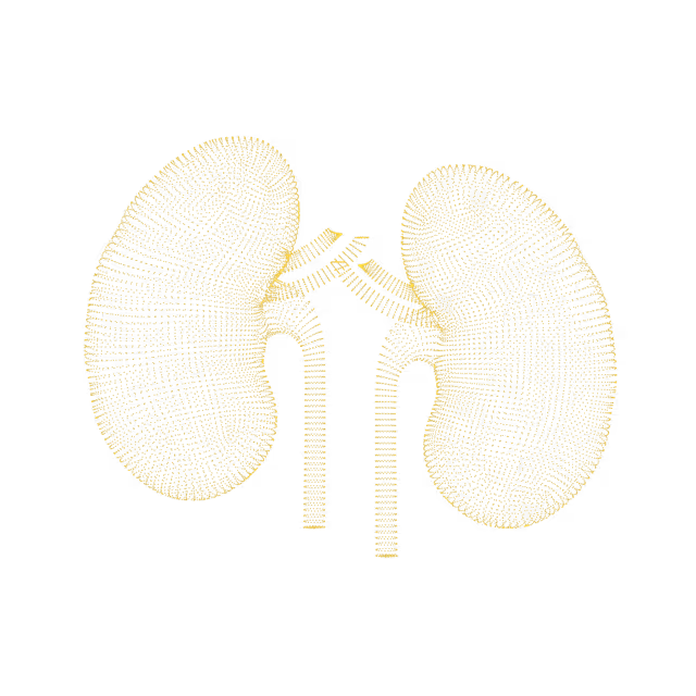
As a father with a family history of cancer, Mike has made it his mission to take control of his health. Mike's Ezra scan found cancer in his kidneys before it was a problem.
"A large part of the credit for this great prognosis goes to early detection: given that the tumor was found so early, it was easier to remove surgically, and any spread is unlikely"
Holly Hollings, 72.
Holly Hollings, two-time cancer survivor, learnt about her early-stage uterus cancer after her Ezra Scan showed an abnormal finding.
“Most people diagnosed with cancer twice can’t say cancer and lucky in the same sentence. I'm so thankful to have caught these cancers early.”
Sam Hires, 72.
Sam's Ezra Scan found Stage 3 kidney cancer - but luckily he found it in time. Thanks to his proactive approach to health Sam will continue living a happy and active life. “I’ve always taken my health seriously so I was shocked that the Ezra scan showed stage 3 kidney tumors on both kidneys. My physician says taking the scan has given me a chance for a longer life.”
Oops! Something went wrong while submitting the form.
PSA density
The PSA (prostate specific antigen) density is an additional tool that may be used to predict the likelihood of prostate cancer. It is the PSA level corrected for the size of the prostate. The PSA density is calculated by dividing the PSA level by the volume of the prostate. Prostate volume is calculated from MRI or transrectal ultrasound measurements. PSA Level ÷ Prostate Volume = Your PSA Density (ng/mL)Generally, if the PSA density is less than 0.15, a prostate biopsy can be reasonably avoided or delayed. A PSA density greater than or equal to 0.20 increases the suspicion of a clinically significant prostate malignancy (cancer).
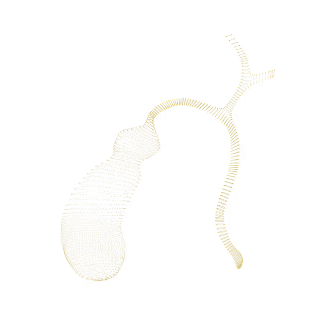
Pelvis
Spermatic cord lipoma
The spermatic cords are part of the male reproductive system, carrying sperm out of the testicles. Lipomatous thickening of the spermatic cords, also known as spermatic cord lipoma or inguinal canal lipoma, is a benign (non-cancerous), fatty growth and demonstrates a characteristic appearance on MRI.
Pelvis
Retractile testicle
The inguinal canal is a passage in the abdominal wall near the groin. In men it serves as a pathway through which the spermatic cords can pass from the abdominal wall to the external genitalia. There is one inguinal canal in each groin. Some individuals have a retractile testicle, in which the testicle is drawn into the inguinal canal by the cremaster muscle reflex. This reflex is elicited when the inner part of the thigh is touched, causing the cremaster muscle to pull up the testicle into the inguinal canal. If the testicle can be manually moved back into the scrotum and stay in the scrotum on its own, it is not considered a serious health condition. Retractile testicles generally do not need to be treated unless you are having testicular discomfort or infertility issues.
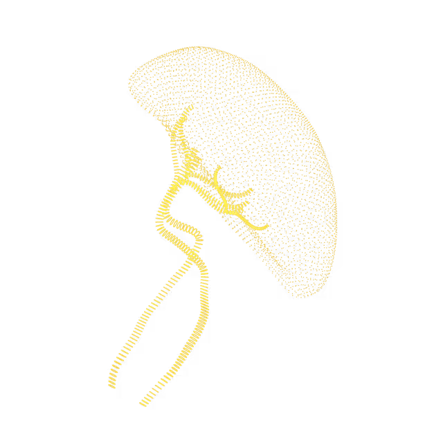
Pelvis
Scrotoliths / scrotal pearl
Scrotoliths, also known as scrotal pearls, are benign (non-cancerous) calcifications (stones) within the scrotum (the small, muscular sac that contains and protects the testicles, blood vessels, and part of the spermatic cord). Although their exact cause is unclear, they are closely associated with chronic microtrauma to the scrotal region (e.g. from mountain biking, horseback riding) which causes damage to the tissues of the scrotum. Other risk factors include twisting of the epididymis (the long coiled tube that is above and behind the testicle and where sperm mature), twisting of the appendix testis (a small portion of normal tissue that is usually located on the upper portion of the testis), and having a hydrocele (a collection of fluid within the scrotum that directly surrounds the testes and the spermatic cord).
Sometimes a testicle is not seen in adults on imaging - a condition called postpubertal cryptorchidism. This could be due to the testicle not forming prior to birth (congenital) or because the testicle never descended into the scrotum and slowly shrank in size (atrophied). Postpubertal cryptorchidism is associated with lower fertility, impaired hormone function and an increased risk of testicular cancer.
Pelvis
Lesions within the vaginal wall (Vaginal wall lesions)
Inclusion cysts are mucus collections that form a sac. Sometimes these cysts can rupture and release blood, known as hemorrhagic cysts. Proteinaceous cyst refers to a cyst with a thicker protein fluid inside. Inclusion cysts can be caused by childbirth or minor physical trauma. They are benign (non-cancerous) and are usually asymptomatic (do not cause symptoms), but sometimes can be the cause of pain or a bothersome feeling of fullness in the vagina.
Pelvis
Epidermal inclusion cyst of the pelvis
Epidermal inclusion cysts are the most common cysts that affect the skin. Most epidermal inclusion cysts form when the cells from the surface of the skin (epidermis) move deeper into the skin and multiply rather than slough off. They often occur in areas where hair follicles have been inflamed and are common in conjunction with acne.Epidermal inclusion cysts can occur anywhere on the body and typically present as skin-colored nodules that are non-cancerous. The size ranges from a few millimeters to several centimeters in diameter. Nodules may remain stable and painless or progressively enlarge. Spontaneous inflammation and rupture can occur, with involvement of surrounding skin.
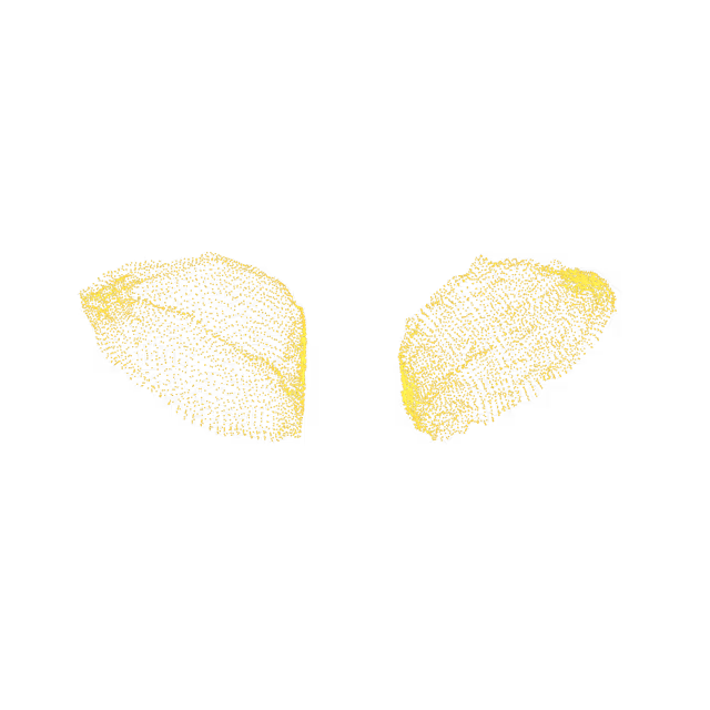
Pelvis
Epididymal cyst (Spermatocele)
An epididymal cyst is a fluid collection in the epididymis, which is a long coiled tube that is above and behind the testicle and where sperm mature. An epididymal cyst may be felt as a soft round mass in this area. A cyst that is larger than 2 cm is called a spermatocele. The exact cause of epididymal cysts is unknown. These cysts usually do not cause symptoms and do not require further monitoring or treatment.
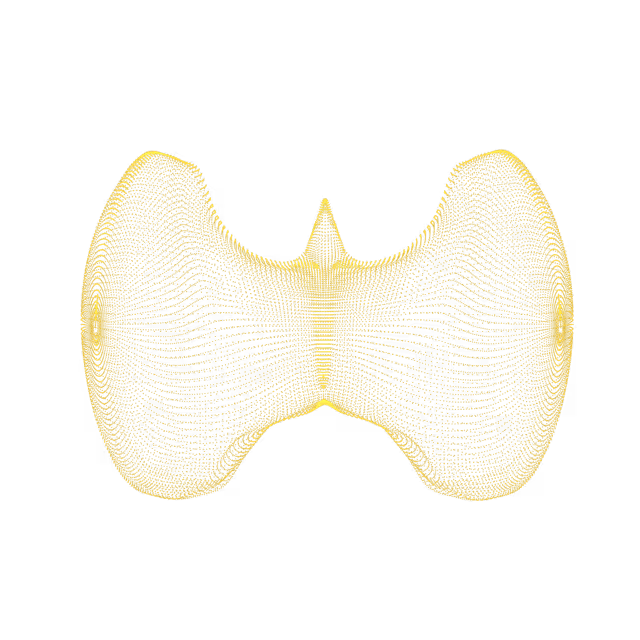
Pelvis
Inflammation of the seminal vesicles
Inflammation of the seminal vesicles is most commonly caused by an infection and is often associated with concurrent infection elsewhere in the male genital tract. It is usually acute, but can be chronic. Risk factors for inflammation of the seminal vesicles include chronic prostatitis (inflammation of the prostate), urethritis (inflammation of the urethra, the tube that carries urine from the bladder out of the body), and acute epididymitis (inflammation of the epididymis, the tube located at the back of the testicles that stores and carries sperm).The spectrum of symptoms of seminal vesicle inflammation is broad and includes ejaculatory dysfunction (e.g. painful ejaculation, decreased ejaculate volume), infertility, pain (e.g. pelvic, flank, abdominal, groin), abdominal swelling, hematuria (blood in the urine) and pneumaturia (the passage of gas or “air” in the urine).
Pelvis
Chronic blood products in the seminal vesicle (hematospermia)
Hematospermia, the term used to describe the presence of blood in the semen, is an uncommon condition. While it is usually alarming to individuals, the condition typically resolves spontaneously. The cause is almost always benign (non-cancerous) and often no clear cause can be identified, which can make it a challenging condition. It can manifest as a single episode or recur over the course of weeks to months. Known causes include, but are not limited to, following radiation treatment for prostate cancer, sexually transmitted diseases, and bleeding from blood thinners. In individuals over 40 with hematospermia, genitourinary cancers (i.e. prostate, seminal vesicles) should be considered, but the rate of such cancers is low, even after long-term follow-up.
We simplify your health insights so you can quickly take action to improve your health. You can view risk levels for each region of your body and book a call with your Ezra Medical Provider for further insights into your body.
See your reports and images in MyEzra
Book a video consultation to dive deeper into your results