We find potential cancer in 6% of our members. 46% find an actionable clinical finding.
Mike Mettler, 45.
As a father with a family history of cancer, Mike has made it his mission to take control of his health. Mike's Ezra scan found cancer in his kidneys before it was a problem.
"A large part of the credit for this great prognosis goes to early detection: given that the tumor was found so early, it was easier to remove surgically, and any spread is unlikely"
Holly Hollings, 72.
Holly Hollings, two-time cancer survivor, learnt about her early-stage uterus cancer after her Ezra Scan showed an abnormal finding.
“Most people diagnosed with cancer twice can’t say cancer and lucky in the same sentence. I'm so thankful to have caught these cancers early.”
Sam Hires, 72.
Sam's Ezra Scan found Stage 3 kidney cancer - but luckily he found it in time. Thanks to his proactive approach to health Sam will continue living a happy and active life. “I’ve always taken my health seriously so I was shocked that the Ezra scan showed stage 3 kidney tumors on both kidneys. My physician says taking the scan has given me a chance for a longer life.”
Oops! Something went wrong while submitting the form.
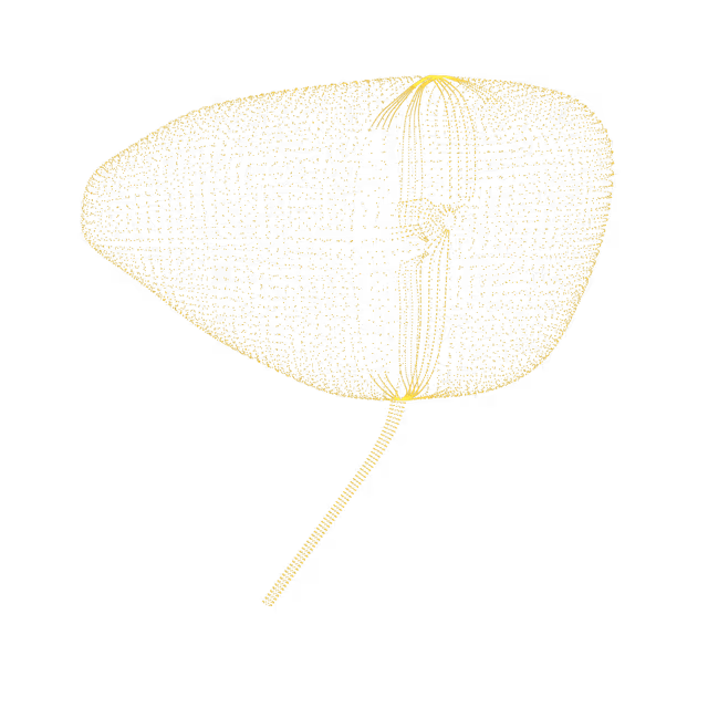
Retroverted and anteflexed uterus
A retroverted uterus is a common condition in which the uterus tilts towards the back rather than to the front. An anteflexed uterus means the top of the uterus (fundus) is pointing forward, which is a normal position.
Pelvis
Subserosal fibroid
Uterine fibroids (also called uterine leiomyomas or myomas) are abnormal growths in the muscle of the uterus. Approximately 80% of women will have fibroids in their lifetime. Subserosal fibroids grow outward from the uterus to the pelvic cavity.
Pelvis
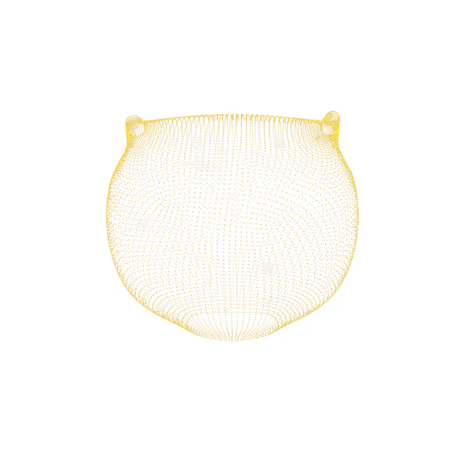
Unicornate uterus
A unicornuate uterus is a rare condition that is present from birth (congenital) that causes an individual to have only half a uterus. Individuals with a unicornuate uterus have one working fallopian tube (instead of two) and a smaller uterine cavity. The body reabsorbs the other half of the uterus. A rudimentary horn is what’s left of the uterine tissue that was absorbed. The rudimentary horn may or may not be connected to the unicornuate uterus. In addition, the rudimentary horn may or may not have functioning endometrial lining/cavity. The condition is usually asymptomatic (does not cause symptoms) but sometimes can be associated with increased risk of adverse pregnancy outcomes (i.e. miscarriages, preterm labor).
Pelvis
Adenomyoma
An adenomyoma (focal adenomyosis) is a non-cancerous abnormal growth where endometrial tissue (the innermost lining layer of the uterus) is inside the uterus muscle, resulting in a mass. Symptoms, if present, may include heavier than normal menstrual bleeding, pain with menstruation, and painful intercourse.
Pelvis
Leiomyosarcoma
Leiomyosarcoma isa rare, malignant (cancerous) tumor that arises from the smooth muscle lining the walls of the uterus. While the exact cause of a uterine leiomyosarcoma is unknown, associated risk factors include long-term use of tamoxifen (five years or more), history of pelvic radiation, and inherited conditions (e.g. childhood retinoblastoma)
Pelvis
Atrophy of uterus and adnexa
Uterine and adnexal (ovaries and fallopian tubes) atrophy is a response to a hypo-estrogen state (low estrogen levels in the body). Atrophy describes when tissue “wastes away” or thins. The most common cause for a hypo-estrogen state is menopause. Other factors that can cause uterine and adnexal atrophy include prolonged oral contraception, ovarian dysfunction and tamoxifen use (estrogen-lowering medication). Atrophy is a normal change to the uterus and adnexa as a woman ages and goes through menopause.
Pelvis
Adnexal scarring
The adnexa describes the region encompassing the ovary and fallopian tube. The adnexa appears scarred, likely consistent with a history of having pelvic surgery.
Pelvis
Salpingo-oophorectomy
The ovary and fallopian tube was not seen on the MRI images which is consistent with a history of surgical removal of your ovary and fallopian tube (salpingo-oophorectomy).
Pelvis
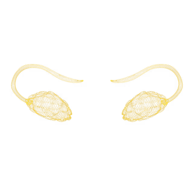
Enlarged ovary with numerous follicles
An enlarged ovary is an ovary that has expanded past its normal size. The cause of this enlargement is usually cyst formation. Other causes include endometriosis, benign tumors and, rarely, ovarian cancer. Each month, the ovaries normally grow cyst-like structures called follicles. These follicles produce the hormones estrogen and progesterone and release an egg during ovulation. Sometimes numerous follicles can be a finding consistent with polycystic ovarian syndrome (PCOS), or it could be from a regular, normal menstrual cycle
Pelvis
Corpus luteal cyst
Each month, the ovaries normally grow cyst-like structures called follicles. These follicles produce the hormones estrogen and progesterone and release an egg during ovulation. After releasing its egg, the follicle is called the corpus luteum. Sometimes, fluid accumulates inside the follicle, causing the corpus luteum to grow into a cyst.
We simplify your health insights so you can quickly take action to improve your health. You can view risk levels for each region of your body and book a call with your Ezra Medical Provider for further insights into your body.
See your reports and images in MyEzra
Book a video consultation to dive deeper into your results