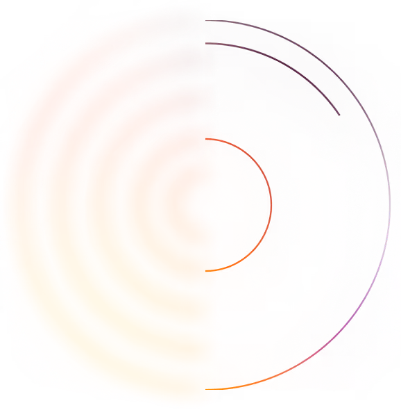
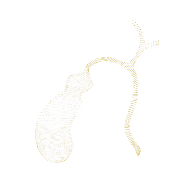
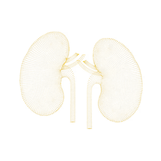
We find potential cancer in 6% of our members. 46% find an actionable clinical finding.
Mike Mettler, 45.
As a father with a family history of cancer, Mike has made it his mission to take control of his health. Mike's Ezra scan found cancer in his kidneys before it was a problem.
"A large part of the credit for this great prognosis goes to early detection: given that the tumor was found so early, it was easier to remove surgically, and any spread is unlikely"
Holly Hollings, 72.
Holly Hollings, two-time cancer survivor, learnt about her early-stage uterus cancer after her Ezra Scan showed an abnormal finding.
“Most people diagnosed with cancer twice can’t say cancer and lucky in the same sentence. I'm so thankful to have caught these cancers early.”
Sam Hires, 72.
Sam's Ezra Scan found Stage 3 kidney cancer - but luckily he found it in time. Thanks to his proactive approach to health Sam will continue living a happy and active life. “I’ve always taken my health seriously so I was shocked that the Ezra scan showed stage 3 kidney tumors on both kidneys. My physician says taking the scan has given me a chance for a longer life.”
Oops! Something went wrong while submitting the form.
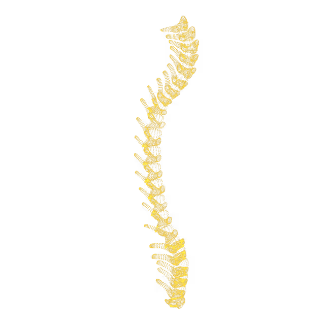
"Butterfly" vertebra
Sometimes during fetal development a vertebra (spine bone) does not fuse together properly, ending slightly wider than a normal vertebra and appearing like a “butterfly” on radiological imaging. This is a normal variant and not cancerous. “Butterfly” vertebra can make the affected area more prone to chronic back pain.
Spine
Vertebral Compression fracture
A vertebral compression deformity (fracture) describes vertical collapse of the spine bone. This is commonly caused by past trauma such as a car accident, sports injury or a hard fall. Symptoms of a subacute compression fracture include back pain, an increase in pain intensity while standing or walking as well as difficulty bending over or sitting.
Spine
Anterolisthesis
Anterolisthesis (also known as spondylolisthesis) is a forward slipping of the vertebra (spinal bone) over the bone below it. A pars defect is a stress fracture of the pars interarticularis, a thin bone connecting two vertebrae. Pars defects are a common injury caused by wear-and-tear and are often seen in those involved in high-risk sports (e.g. gymnastics, football, wrestling, weight lifting).
Spine
Bone cyst
Bone cysts are benign (non-cancerous) lesions that develop inside a bone. Bone cysts usually do not cause any symptoms. However, sometimes they can expand in size and cause symptoms of pain or numbness when the adjacent nerve is compressed.
Spine
Bone island of the spine
A bone island, also known as an enostosis, is a common incidental finding (found in passing when looking for something else) of a small area of dense bone inside the spongy part of the bone. Enostoses are common, present in up to 14% of individuals.The cause of bone islands is not known. They are most likely congenital (present from birth) or developmental in nature and are thought to represent either hamartomatous lesions (non-cancerous tumorlike malformations made up of an abnormal mixture of cells and tissues found in areas of the body where growth occurs) or failure of osteoclastic activity (the body's process of breaking down bone in order to build it up again) during bone remodeling. Bone islands are usually considered benign (non-cancerous), stable, nonprogressive lesions, with a preference for the long bones and the pelvis. They typically present without symptoms, do not cause pain and usually do not grow larger. Although bone islands have a characteristic appearance in the vast majority of cases, larger lesions may sometimes pose a diagnostic dilemma, particularly in the setting of known malignancy.
Spine
Congenital spinal canal stenosis
Congenital (from birth) spinal canal stenosis is when the space where the spinal cord runs through the spine is narrower than normal. Symptoms, if present, can include pain, numbness, tingling and/or weakness which could radiate into the extremities (arms or legs).
Spine
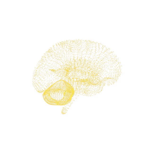
Centrum semiovale white matter plaques
The centrum semiovale is the white matter oval shaped core of the cerebral hemispheres (the left and right side of the brain). White matter is the brain tissue that contains nerve fibers and serves as the connection to other parts of the brain. White matter hyperintensities are common changes seen on MRI in asymptomatic individuals (those with no symptoms), and their prevalence increases with age, to nearly 100% in those older than 90 years. The possible causes of white matter hyperintensities include chronic microvascular ischemic changes, vasculitis (blood vessel inflammation), migraine, Lyme disease or demyelinating disease.
Brain
Indeterminate bone lesion
A lesion is an abnormality seen on an imaging test. A bone lesion may involve small to large areas of your bone(s) and the severity of the underlying condition may range from relatively minor to life-threatening. These lesions will need further work up to for a diagnosis.
Spine
Scattered foci of T2 hyperintensities involving the supratentorial white matter
The supratentorial area is the upper part of the brain and the infratentorial area is the lower back part of the brain. White matter is the brain tissue that contains nerve fibers and serves as the connection to other parts of the brain. White matter hyperintensities are areas with high water or protein content that show up bright and are common changes seen on MRI in asymptomatic individuals (those with no symptoms). Their prevalence increases with age, to nearly 100% in those older than 90 years. The possible causes of white matter hyperintensities include chronic microvascular ischemic changes, vasculitis (blood vessel inflammation), migraine, Lyme disease or (less likely) demyelinating disease.
Brain
Nonspecific subcortical white matter T2 hyperintensities
White matter is the brain tissue that contains nerve fibers and serves as the connection to other parts of the brain. It is found in the subcortical (deeper) tissues of the brain. Nonspecific (meaning it is difficult to say what caused it) T2 hyperintensities describe areas with high water or protein content that show up “bright” (hyperintense) on certain MRI sequences. White matter hyperintensities are common changes seen on MRI in asymptomatic individuals (those with no symptoms), and their prevalence increases with age, to nearly 100% in those older than 90 years. They might be simply a benign (non-cancerous) marker of aging or they could be a marker of small vessel disease which is associated with cognition and stroke. Modifiable risk factors of small vessel disease include smoking, high blood pressure, poor cholesterol levels, carotid artery disease, and atrial fibrillation.
We simplify your health insights so you can quickly take action to improve your health. You can view risk levels for each region of your body and book a call with your Ezra Medical Provider for further insights into your body.
See your reports and images in MyEzra
Book a video consultation to dive deeper into your results