Your vital organs—screened
Scan your body for potential cancer and 500+ conditions in up to 13 organs.


Our scan is designed to


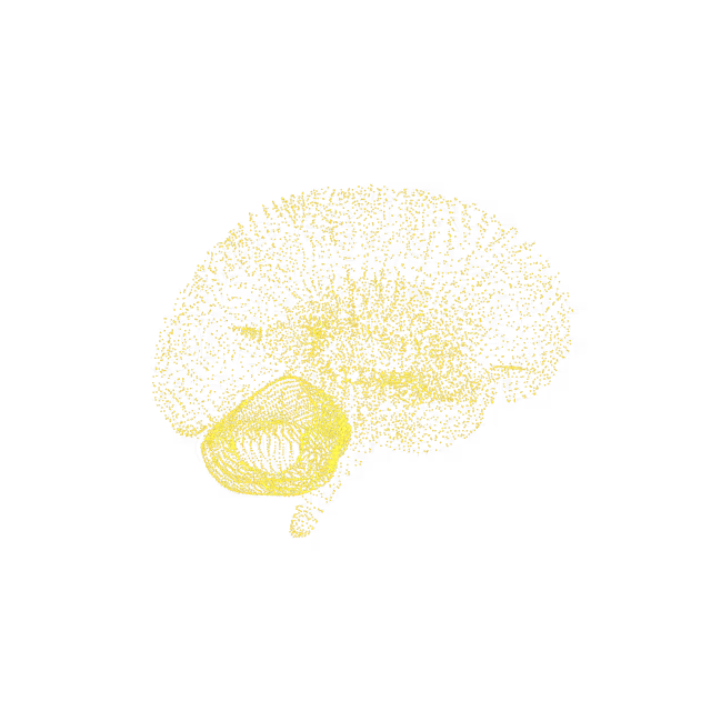

Encephalomalacia describes softening of brain tissue, usually from a past stroke, trauma or infection, or could be congenital (from birth). The most common cause of encephalomalacia is stroke, which has the same major risk factors that lead to heart disease (high blood pressure, poor cholesterol levels, diabetes and smoking).
A localized area (i.e. focus) that appears abnormal on MRI brain images. This finding is nonspecific, meaning it is difficult to say what caused it.
Glial cells are the support and insulating cells for neurons (which conduct electrical impulses in the central nervous system). Gliosis is when the body creates more or larger glial cells in reaction to some injury to the brain or spinal cord. Any injury (such as physical trauma) or inflammatory process (such as an autoimmune condition) of the central nervous system can cause gliosis; however, the role of the process is unknown.
The brain and spinal cord are covered by three protective membrane-linings called meninges. The arachnoid membrane, named for its spider web-like appearance, is the thin middle layer. Blood and fluid are drained from the brain through sinuses. The superior sagittal sinus runs midline in the brain and is the largest dural venous sinus (a group of blood channels that drains blood from the brain).Arachnoid granulations are projections of the arachnoid membrane (i.e. villi) into the dural sinuses. Arachnoid granulations act as one-way valves, allowing cerebrospinal fluid to pass from the subarachnoid space into the venous (blood) circulation. They increase in size and number with age and are seen in approximately two-thirds of the population.
A lacunar stroke (infarct) is when blood flow going to the small arteries deep inside the brain become blocked. The major risk factor for getting lacunar strokes is chronic high blood pressure, which can cause the small arteries to narrow over time; other modifiable risk factors include poor cholesterol levels, diabetes and smoking.If there is a new stroke, think “FAST” - Facial drooping, Arm weakness, Speech difficulties, Time is of the essence - call 911. Treatment is focused on restoring blood flow to the brain.
A glioma is an abnormal growth of cells (tumor) that forms from glial cells found in the brain and spinal cord. Glial cells provide the structural backbone of the brain and support the function of neurons (nerve cells). Brain tumors are classified as primary, those that arise in the brain, or secondary, those that have spread to the brain from another part of the body. In the United States, about 24,000 people per year are diagnosed with a primary brain tumor. Most primary brain tumors in adults have no clear risk factors identified. Brain tumors can produce symptoms due to local brain invasion, compression of healthy brain structures and by increasing pressure within the brain (increased intracranial pressure). Symptoms vary based on what parts of the brain are involved.
Gyri are ridges on the surface of the brain. There is a mass effect or compressing/pushing of the adjacent gyri from the arachnoid cyst mentioned above.Potential Cancer
Brain tissue tends to shrink (atrophy) at the rate of about 0.2% per year from the age of 30 and then accelerates after the age of 60, due to genetic, environmental, and lifestyle factors. Volume loss which is out of proportion to age may represent a pathologic process or risk factor for future decline in cognition. There are currently no established guidelines for investigating or monitoring this condition; however, you could consider repeat MRIs to monitor progression or stability of this finding.
Microangiopathy is a term that is used to describe changes to the small blood vessels in the brain. The cause is not completely understood, but could be the result of plaque buildup and hardening (atherosclerosis) of the small blood vessels nourishing the brain. This is the same process that can narrow and damage heart blood vessels.
The cerebellum is the area at the back and bottom of the brain, behind the brainstem (where the spinal cord meets the brain). It receives information from the sensory systems, the spinal cord, and other parts of the brain and then regulates motor movements. The cerebellum coordinates voluntary movements such as posture, balance, coordination, and speech, resulting in smooth and balanced muscular activity. It is also important for learning motor behaviors and coordination of eye movements. Diffuse atrophy of the cerebellum refers to a progressive and irreversible reduction in cerebellar volume. It is found in a wide variety of clinical scenarios. It can also result from a variety of causes including drugs (alcohol and/or certain medications) and neurodegenerative diseases.


© 2025 Ezra