We find potential cancer in 6% of our members. 46% find an actionable clinical finding.
Mike Mettler, 45.
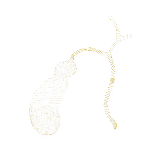
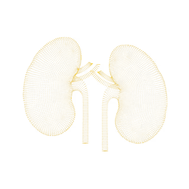
As a father with a family history of cancer, Mike has made it his mission to take control of his health. Mike's Ezra scan found cancer in his kidneys before it was a problem.
"A large part of the credit for this great prognosis goes to early detection: given that the tumor was found so early, it was easier to remove surgically, and any spread is unlikely"
Holly Hollings, 72.
Holly Hollings, two-time cancer survivor, learnt about her early-stage uterus cancer after her Ezra Scan showed an abnormal finding.
“Most people diagnosed with cancer twice can’t say cancer and lucky in the same sentence. I'm so thankful to have caught these cancers early.”
Sam Hires, 72.
Sam's Ezra Scan found Stage 3 kidney cancer - but luckily he found it in time. Thanks to his proactive approach to health Sam will continue living a happy and active life. “I’ve always taken my health seriously so I was shocked that the Ezra scan showed stage 3 kidney tumors on both kidneys. My physician says taking the scan has given me a chance for a longer life.”
Oops! Something went wrong while submitting the form.
Acoustic schwannoma
An acoustic schwannoma (also known as acoustic neuroma or vestibular neuroma) is a benign tumor that develops around a nerve in the inner ear. This tumor grows around and compresses either the auditory (cochlear -for hearing) or vestibular (for balance) nerves leading from your ear to the brain.
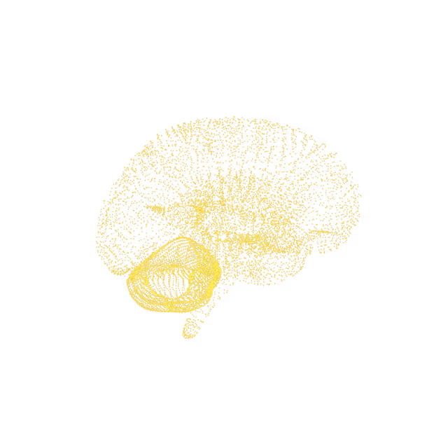
Brain
Demyelinating disease
A demyelinating disease is any condition that causes damage to the protective covering (myelin sheath) that surrounds nerve fibers in your brain, the nerves leading to the eyes (optic nerves) and spinal cord. When the myelin sheath is damaged, nerve impulses slow or even stop, causing neurological problems.
Brain
Indeterminate brain lesion
A brain lesion is an abnormality seen on a brain-imaging test. A brain lesion may involve small to large areas of your brain, and the severity of the underlying condition may range from relatively minor to life-threatening. These lesions will need further work up to for a diagnosis.
Brain
Pulmonary/lung nodule
A pulmonary or lung nodule is a “spot” seen on chest imaging. This can be caused by infection, scarring or lung cancer.
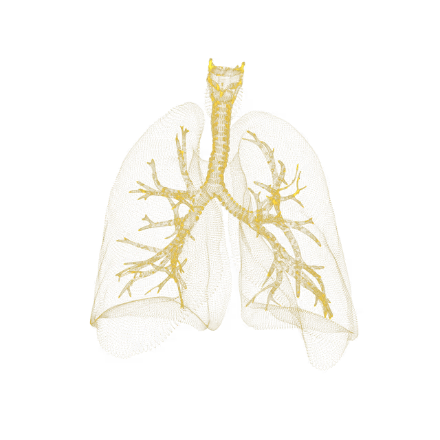
Chest/Lung (CT)
Lung cancer
Cancer that forms in tissues of the lung, usually in the cells lining air passages. The two main types are small cell lung cancer and non-small cell lung cancer. These types are diagnosed based on how the cells look under a microscope.
Chest/Lung (CT)
Pituitary tumor
Pituitary tumors are unusual growths that develop in the pituitary gland. This gland is an organ about the size of a pea. It's located behind the nose at the base of the brain. Some of these tumors cause the pituitary gland to make too much of certain hormones that control important body functions. Others can cause the pituitary gland to make too little of those hormones. Most pituitary tumors are benign.
Brain
Intrathoracic endometriosis
Thoracic endometriosis is a rare condition that happens when endometriosis patches grow on or around the lungs. This can cause shortness of breath, chest pain, cough, and in some cases, a collapsed lung.
Chest/Lung (CT)
Breast calcifications
Breast calcifications (hard, dense, calcium deposits within the breast) appear as white spots on imaging.
Chest/Lung (CT)
Enlarged heart
An enlarged heart (cardiomegaly) refers to a heart that is bigger than normal. This condition may be seen on imaging (e.g. MRI, CT) in individuals with an underlying condition (e.g. high blood pressure, coronary artery disease, inherited disorders, cardiomyopathy) that causes the heart muscle to become thicker or a condition that causes one of the chambers of the heart to dilate, making the heart larger.
Chest/Lung (CT)
Left vertricular hypertrophy
The heart has 4 main chambers - 2 atria, one that receives blood from the body and another from the lungs, and 2 ventricles, one which pumps blood to the body and the other to the lungs. The left ventricle appears enlarged (hypertrophy). Causes of left ventricle enlargement include: “normal” athlete’s heart, high blood pressure, aortic valve stenosis and hypertrophic cardiomyopathy.
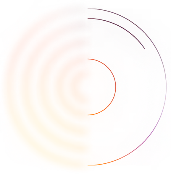
We simplify your health insights so you can quickly take action to improve your health. You can view risk levels for each region of your body and book a call with your Ezra Medical Provider for further insights into your body.
See your reports and images in MyEzra
Book a video consultation to dive deeper into your results