Introduction
Cysts are sac-like structures filled with fluid, air, or other materials, and they can appear in various parts of the body, including skin, organs, and tissues. While most cysts are benign, some can be early warning signs of cancer, making it essential to monitor and evaluate any unusual lumps or growths in the body1. Early detection and screening are important for managing cysts effectively. Imaging technologies, including ultrasound, computed tomography (CT), and magnetic resonance imaging (MRI), play a vital role in identifying and characterizing cysts.
What are Cysts?
Cysts are closed sac-like structures that can form in various parts of the body, containing fluid, semi-solid material, or gas. These abnormal formations have distinct membranes or walls that separate them from surrounding tissues2. Cysts can develop in almost any type of body tissue and vary in size from microscopic to large structures that might displace internal organs3. They can occur for various reasons, including infections, tumors, parasites, or injuries.
Types of Cysts and Cancer Risks
Most cysts are benign and harmless, often resolving independently without treatment. However, some cysts have the potential to become malignant. Certain types of cysts, such as cystadenomas and dermoid cysts, carry a higher risk of being cancerous, particularly in postmenopausal women or those with a family history of ovarian cancer4. Cancer-related cysts may form as a defense mechanism following mutations that lead to uncontrolled cellular division5. While the risk of cancer in those with complex ovarian cysts is generally low, it’s higher than in simple cysts6.
How Do You Detect Cancer in Cysts?
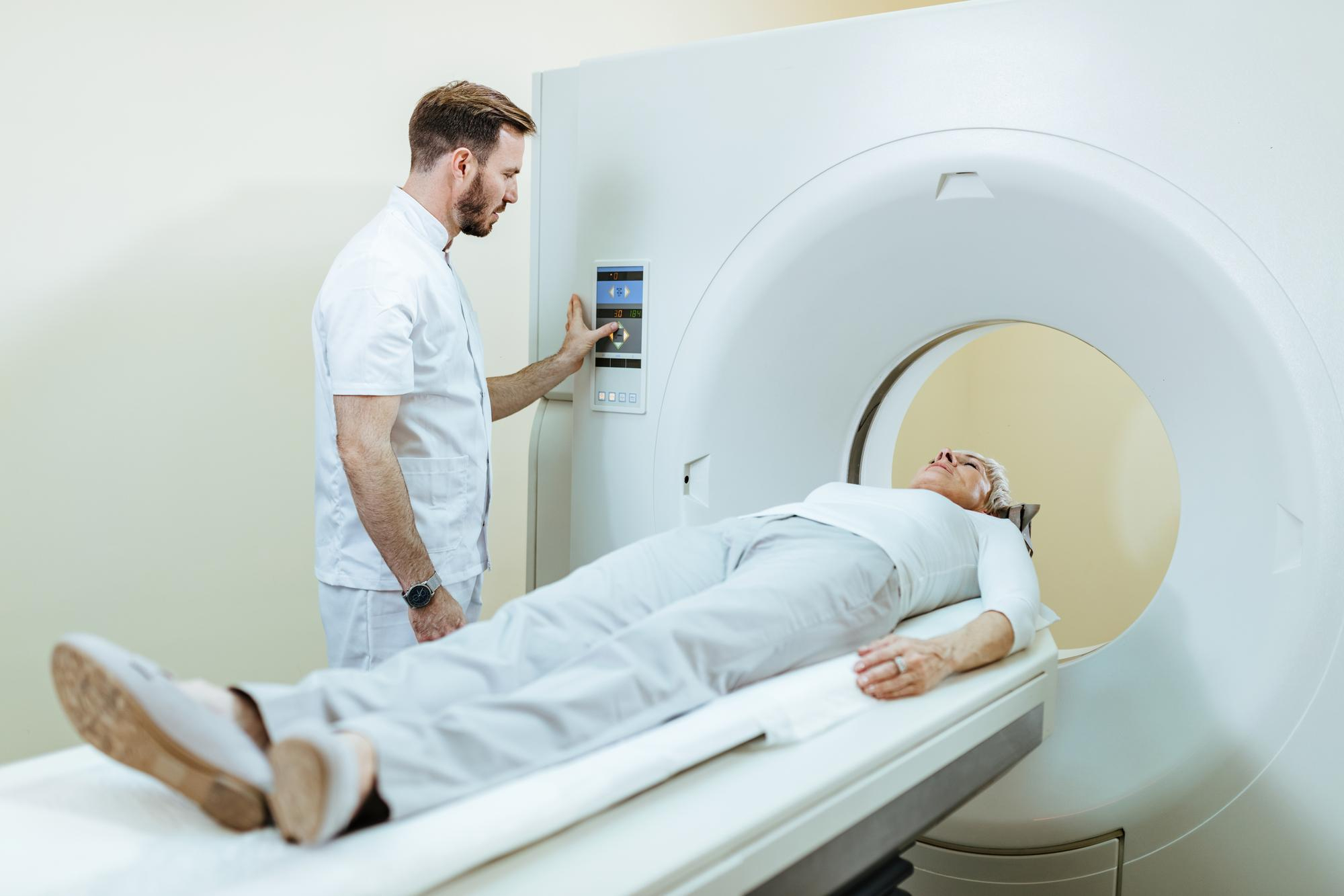
Imaging Techniques
CT scans are crucial in detecting cancer in cysts by providing detailed information about their size and structure7. They offer high precision in capturing cyst details and potential cancer markers, allowing for accurate differentiation between benign and malignant formations8.
MRI scans are particularly effective in highlighting subtle details of cysts, including components that suggest a higher risk of cancer9. Their superior soft-tissue contrast allows for a more nuanced evaluation of cyst characteristics, making them especially useful for assessing pancreatic cysts and masses around the uterus (adnexal masses)10.
Ultrasound can detect features indicative of malignancy, such as irregular solid components, papillary projections, and increased vascularity11. Ultrasound is often the first-line imaging method for young, pregnant, and breastfeeding patients with clinical symptoms.
Other Diagnostic Methods
Biopsies are typically used to confirm malignancy in cysts. This procedure involves removing a sample of the affected tissue or, in some cases, the entire suspicious area for microscopic examination12. Blood tests are also conducted for tumor markers. These play a significant role in cyst evaluation, particularly for ovarian cysts13.
Symptoms of Cancerous Cysts
Symptoms of cancerous cysts can be similar to those of benign cysts, making it important to understand the key differences between them. While most cysts are harmless, certain signs may indicate a higher risk of malignancy.
Common Symptoms
Pain is a frequent symptom of both benign and cancerous cysts. For ovarian cysts, this can manifest as pelvic pain ranging from a dull sensation to sharp, severe pain14. Changes in size or shape, particularly rapid growth, can be a concerning sign. Systemic symptoms like fatigue, unexplained weight loss, or fever may also occur with cancerous cysts15. In the case of ovarian cysts, women might experience bloating, feeling full quickly after eating, and changes in urinary habits.
Red Flags
Persistent growth and irregular borders on imaging are potential red flags for cancerous cysts16,17. Solid components within a cyst may indicate a higher risk of malignancy. Other concerning signs include changes in color, bleeding or oozing, and redness or swelling around the cyst.


