Neck cancer refers to cancers that affect various structures in the head and neck, including the oral cavity, pharynx, and larynx1. They can impact vital functions like speech and swallowing. Early detection is important for effective treatment, and CT scans are a key tool for providing detailed images of internal structures.
What is Neck Cancer?
Neck cancer is an umbrella term for several types of cancers that affect the structures in the neck region. This includes malignancies such as salivary gland cancer, carotid body tumors, thyroid cancer, lymphoma, and squamous cell carcinoma2.
The symptoms can vary depending on the type and location of the cancer. Many patients notice a persistent lump or swelling in the neck, a sore throat that does not improve over time, difficulty swallowing, changes in the voice, ear pain, or unexplained weight loss3.
Risk factors include:
- Tobacco use4.
- Alcohol consumptionr5.
- Human Papillomavirus (HPV) infection6.
- Environmental exposures7.
- Poor diet8.
- Family history9.
It should be noted that having one or more risk factors doesn’t necessarily mean a person will develop neck cancer.

CT Scans vs. Other Imaging Techniques
CT scans are valuable for cancer detection in the neck by providing cross-sectional images that reveal internal abnormalities. However, they do involve exposure to ionizing radiation. For every 100,000 neck CT scans, estimates suggest a small increase in subsequent cancer cases (ranging from a few thyroid cancers to higher numbers in specific populations)10.
Other imaging options include:
- Ultrasound: Often more sensitive than CT in detecting primary tumors (74 percent vs. 68 percent) and recurrent cancers (67 percent vs 63 percent)11. However, ultrasound also had a high rate of false-positive results12.
- MRI: Excels at soft tissue characterization without the risk of ionizing radiation, making it ideal for visualizing soft tissue tumors13. CT may still be used for evaluating bone involvement or when rapid imaging is needed due to its faster scan time.
- PET/CT: Merges metabolic and anatomical imaging, offering high sensitivity (up to 96.4 percent) and specificity (around 86.4 percent) for detecting lymph node metastases14.
Even with advanced techniques, diagnostic errors occur. For instance, a study found that 4 percent of cases had earlier signs of head and neck cancer on CT or MRI that were missed due to image interpretation errors15.
Cancer Detection in the Neck via CT Scan
CT scans detect neck cancers by revealing differences in tissue density, highlighting abnormal growths. For lymph nodes, CT may show enlarged or irregularly shaped nodes, potentially with central low-density areas suggesting necrosis when contrast-enhanced16. Thyroid nodules may exhibit calcifications or irregular margins, with papillary carcinomas appearing as hypodense (darker) areas with microcalcifications17.
In salivary glands, benign tumors typically have uniform density and well-defined borders, while malignant tumors often show irregular margins and varied contrast enhancement18. Squamous cell carcinoma usually appears as an irregular, infiltrative mass with uneven enhancement and low-density areas indicating central necrosis. Invasion into adjacent tissues is a key marker of malignancy20.
Types of CT Scans for Neck Cancer Detection
A number of CT techniques are used for cancer detection in the neck.
In some cases, contrast will be used to help visualize structures and blood vessels. Intravenous contrast material highlights blood vessels and enhances soft tissue visualization, making tumors more apparent21.
Several advanced CT techniques also enhance neck cancer detection and characterization.
- Dual-energy CT: Provides additional data on tissue composition22.
- Dental cone beam CT: Generates 3D images useful for identifying oral cancers3.
- CT of the sinuses: Used to evaluate paranasal sinus cavities for cancers in the nasal region23.
- Volumetric modulated arc radiotherapy (VMAT) planning: uses CT scans to plan precise radiation therapy24.
These advanced techniques not only improve cancer detection but also aid in treatment planning and monitoring.
Symptoms That Lead to Neck Cancer Screening
While neck cancer symptoms can mimic benign conditions, persistent or multiple symptoms should prompt further evaluation. Key warning signs can include:
Swelling and Lumps: Persistent neck or oral lumps (lasting more than three weeks) and swollen lymph nodes25.
Sore Throat: sore throat that does not improve after 3–4 weeks26.
Voice Changes: Persistent hoarseness or difficulty speaking27.
Oral Abnormalities: Non-healing mouth ulcers, red or white patches, or sores lasting several weeks28.
Additional Warning Signs: Pain when chewing or swallowing, unexplained pain in the face, jaw, or throat, earaches, nasal blockage or nosebleeds, loose teeth, and unintended weight loss
How Early Cancer Detection in the Neck Improves Outcomes
Early detection of neck cancer significantly improves treatment outcomes and survival rates, with early-stage (I or II) survival exceeding 80 percent29. It allows for localized, less invasive treatments that preserve vital functions like speech and swallowing. Early diagnosis enhances surgical planning and enables precise radiation therapy, reducing the need for extensive surgery and minimizing complications.
Additionally, it is cost-effective, preventing the high expenses associated with advanced-stage treatments and lowering healthcare burdens30. Screening programs targeting high-risk populations further improve early detection rates, offering substantial clinical and economic benefits while increasing the likelihood of curative interventions.
Success Rates of CT Scans in Neck Cancer Detection
The success rates of CT scans can vary depending on a number of factors.
CT scans show a sensitivity of 68 percent and a specificity of 69 percent for primary tumor detection11. For recurrent carcinomas, they have a sensitivity of 63 percent and a specificity of 80 percent. Another study using state-of-the-art CT technology reported improved results, with a sensitivity of 83 percent and a specificity of 93 percent for primary tumor detection31.
However, the issue of false positives remains significant; for example, one study reported a specificity as low as 24 percent in post-treatment CT scans, leading to unnecessary invasive procedures and, in about half of those cases, serious complications32.
Preparing for a Neck CT Scan
Proper preparation is essential for a successful CT scan.
You may be asked to fast for four hours before your appointment, although clear fluids and medications are allowed; diabetic patients can have a light snack but should avoid large meals33. If contrast material is required, you might drink a special solution or receive an IV injection21. Always inform your healthcare provider of any allergies, especially to iodine or contrast dyes, and notify them if you’re pregnant or taking medications like metformin, which might need to be paused34.
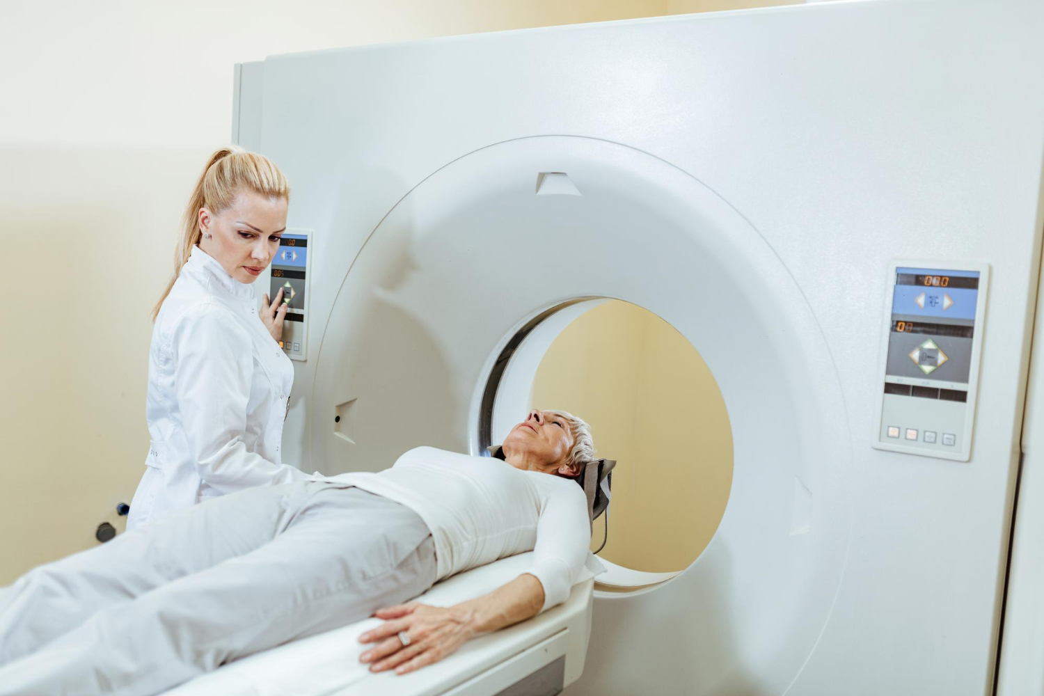
The scan usually lasts 15 to 30 minutes, with the actual imaging taking 5-10 minutes. You will lie on a table that slowly slides into a doughnut-shaped scanner while you remain still. For neck scans, your head will be positioned on a special headrest with small pads placed around it35. You’ll be asked to remain still and be given breathing instructions or asked not to swallow during the scan.
Risks and Limitations of CT Scans
While CT scans are invaluable in the detection and management of neck cancer, they do have some limitations and risks. One of the primary concerns is exposure to ionizing radiation, which can potentially increase the risk of cancer over time by damaging living tissues36. To minimize this risk, low-dose CT (LDCT) protocols have been developed, which significantly reduce radiation exposure while still providing diagnostic-quality images.
The use of contrast agents, though beneficial for image clarity, may pose a risk for patients with allergies or impaired kidney function, prompting the consideration of alternative imaging methods such as MRI or ultrasound37.
CT scans can be limited in detecting very small tumors or early-stage cancers38. However, it remains very important for neck cancer detection when used appropriately.
Next Steps After CT Scan Results
The next steps after a CT scan reveal an abnormality depend on the findings. If a suspicious mass or enlarged lymph nodes are detected, a fine-needle aspiration (FNA) biopsy is often performed to confirm the diagnosis39. Additional imaging, such as MRI or PET/CT, may be needed to determine the extent of cancer, especially for tumors in areas like the throat3,40. If cancer is confirmed, treatment options like surgery, chemotherapy, or radiation are discussed. For negative CT scans but persistent symptoms, follow-up tests or close monitoring may be recommended. Routine imaging is common for patients in remission
Cost and Accessibility of Neck Cancer CT Scans
The cost and accessibility of neck cancer CT scans can vary significantly.
The cost of CT scans for neck cancer can vary considerably depending on factors such as geographic location and insurance coverage. Prices may range from approximately $1,000 to $7,60041.
Insurance status plays a significant role in accessibility; patients with Medicaid or no insurance may face barriers to timely imaging and, as a result, may be more likely to present with advanced-stage tumors compared to those with private insurance.
Latest Advances in Neck Cancer Detection Using CT
Recent advancements in CT technology and related imaging techniques have further improved the detection of neck cancer. The latest generation of digital PET/CT scanners has shown exceptional performance, reporting high sensitivity and specificity for detecting cervical lymph node metastases14.
In addition, integrating artificial intelligence and deep learning into the analysis of CT scans is transforming the field. AI-based algorithms applied to pre-treatment CT scans can predict extranodal cancer spread, accurately segment primary tumors and lymph nodes, and even identify organs at risk during radiotherapy planning42.
Frequently Asked Questions
How effective are CT scans in detecting neck cancer?
CT scans are moderately effective for detecting neck cancer, particularly in assessing tumor size and lymph node involvement. However, they are limited in detecting small tumors and distinguishing post-treatment changes from recurrent disease. Therefore, CT scans are often used with other imaging techniques, such as PET/CT or MRI, for improved accuracy in diagnosis and staging.
What symptoms may indicate the need for a CT scan of the neck?
Swelling and lumps, sore throat, voice changes, oral abnormalities, pain when chewing or swallowing, unexplained face pain, earaches, nasal blockage or nosebleeds, loose teeth, and unintended weight loss are symptoms that might indicate the need for a CT.
How long does a neck CT take?
A neck CT scan typically takes between 10 to 30 minutes. The actual scanning time is usually around 3-5 minutes, but the entire procedure can take longer if contrast is used.
Are CT scans safe for cancer detection?
CT scans are generally considered safe for cancer detection, with the benefits outweighing the small potential risks for most patients. However, there is a slight increase in cancer risk associated with radiation exposure from CT scans.
Can CT scans detect all types of neck cancer?
CT scans are not capable of detecting all types of neck cancer. They are effective for detecting larger tumors, assessing lymph node involvement, and staging certain cancers, like squamous cell carcinoma, but they may miss small or superficial tumors, early-stage cancers, or those with low contrast in soft tissues.
What happens if a tumor is found during a CT scan?
A radiologist will analyze the images and report their findings to the patient's doctor, who will discuss the results and recommend further steps, such as additional imaging or a biopsy. These subsequent tests are crucial to determine whether the tumor is cancerous and to develop an appropriate treatment plan.
Conclusion
CT scans are a cornerstone in the detection, diagnosis, and management of neck cancer, providing detailed imaging that is essential for early tumor identification and precise staging. They play a critical role in guiding treatment decisions and monitoring disease progression, ensuring that therapy is both timely and targeted. Although there are minor risks, the benefits of using CT scans in clinical practice far outweigh these limitations. Ultimately, CT scans improve patient outcomes by facilitating early detection and informed, effective management of neck cancer.
If you are experiencing symptoms or have risk factors for neck cancer, why not book an Ezra Full-Body Plus scan? We combine MRI with LDCT to catch potential cancer earlier, leveraging AI through the screening process to make it more efficient, affordable, and faster.


