Stomach cancer, or gastric cancer, is a malignant tumor in the stomach lining1. Early detection of stomach cancer is crucial for improving survival rates and treatment outcomes, with a significant increase in the 5-year survival rate when diagnosed early2. Imaging technology, particularly computed tomography (CT) scans, plays an important role in identifying stomach abnormalities. CT scans provide detailed cross-sectional images, helping to detect suspicious areas, confirm cancer locations, guide biopsies, and determine treatment options3. They are highly effective for advanced gastric cancer and, with recent advancements, are improving in detecting early-stage cancers as well4.
Understanding Stomach Cancer
As mentioned above, stomach cancer is a malignant tumor that develops in the stomach lining. It occurs when cells in any part of the stomach grow and divide abnormally, forming a mass or tumor5. Most stomach cancers begin in the glandular tissue on the stomach’s inner surface, resulting in adenocarcinomas.
Common symptoms of stomach cancer include6:
Difficulty swallowing (dysphagia).
Unexplained weight loss.
Abdominal pain.
Persistent indigestion (dyspepsia).
Feeling full after eating small amounts.
Loss of appetite.
Nausea and vomiting.
Dark stools (potentially indicating blood).
Risk factors for stomach cancer include:
Being male (men are twice as likely to develop stomach cancer).
Age over 60.
Infection with Helicobacter pylori bacteria.
A diet high in smoked, pickled, and salted foods.
Smoking tobacco.
Alcohol consumption.
Obesity.
Family history of stomach cancer.

When diagnosed at an early stage, the 5-year survival rate for stomach cancer can be as high as 92.6 percent after endoscopic resection7. In contrast, advanced gastric cancer has a poorer prognosis, with overall 5-year survival rates of around 25 percent in Western countries8.
What is a CT Scan?
CT scans are a valuable diagnostic tool in the detection and evaluation of ovarian cancer. They use X-ray technology to create detailed cross-sectional images of the body, providing more information than traditional X-rays9.
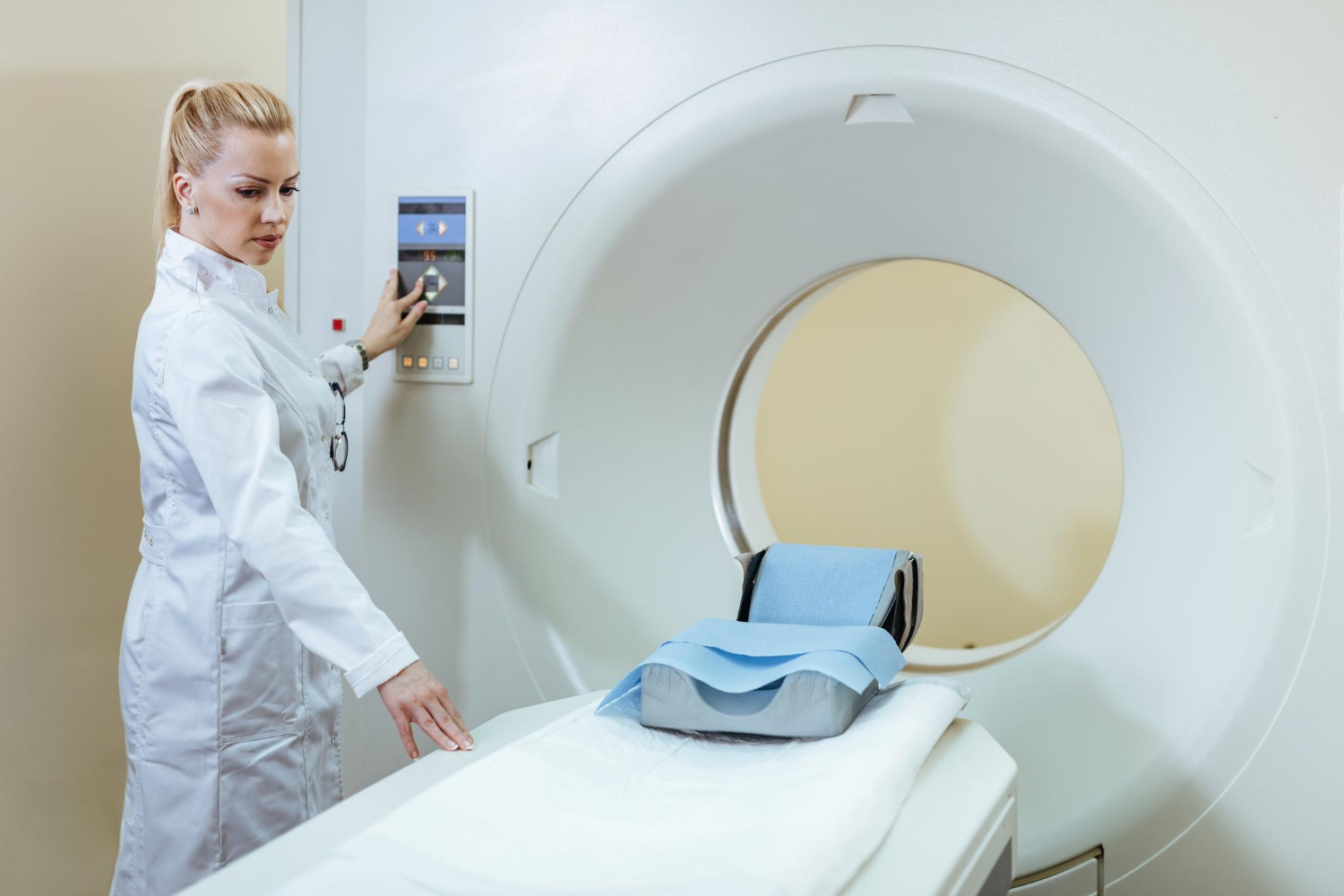
CT scans work by rotating an X-ray beam around the patient’s body, taking multiple images from different angles10. For stomach imaging, the patient typically lies on a narrow table that moves through a donut-shaped machine called a gantry. As the X-ray tube and detectors rotate around the patient, they capture data that is then processed by a computer to generate detailed cross-sectional images of the abdominal area, including the stomach11.
The non-invasive nature of CT scans makes them a preferred choice for many diagnostic purposes. Unlike exploratory surgery, CT scans can provide detailed internal images without the need for incisions or insertions into the body12. This imaging technique is painless and typically takes only a few minutes to half an hour, depending on the area being scanned13. For abdominal CT scans, which include imaging of the stomach, the actual scanning time is usually around 10 minutes.
CT scans are particularly valuable for diagnosing various conditions affecting the stomach and other abdominal organs, such as infections, inflammatory diseases, cancers, and injuries11. They can also be used to guide biopsies, plan treatments, and monitor the effectiveness of ongoing therapies.
How CT Scans Detect Stomach Cancer
CT scans play an important role in detecting abnormalities in the stomach that might indicate cancer. The process involves3:
Image acquisition: The patient lies on a table that moves through the CT machine. The machine then captures multiple cross-sectional images of the abdominal area11.
Contrast enhancement: Patients may drink oral contrast or receive intravenous contrast to improve the visibility of the stomach and surrounding structures.
Image reconstruction: A computer processes the data to create detailed 3D images of the stomach and nearby organs.
Image analysis: Radiologists examine the images for specific features that might indicate stomach cancer.
Tumor Characteristics
Larger tumors are more easily detected and may indicate a higher risk of malignancy. CT scans can confirm the precise location of the tumor within the stomach14. Tumors may appear as:
Endoluminal: Refers to tumors that grow inward, towards the hollow space inside the stomach. These tumors tend to protrude into the stomach cavity, potentially causing obstruction of affecting the stomach’s normal function14.
Exophytic: The tumor grows outward from the stomach wall. These tumors expand towards the outer surface of the stomach, potentially affecting nearby organs or structures as they grow larger15.
Mixed: This type of tumor exhibits both endoluminal and exophytic growth characteristics. It may grow both inward into the stomach cavity and outward from the stomach wall16.
Further research has identified two main types of growth patterns in early stomach cancer: the superficially spreading (Super) type and the penetrating (Pen) type17,18. The Pen type, particularly a subtype called Pen A, which grows expansively and destroys the muscularis mucosae (a layer of the stomach wall), is associated with a less favorable prognosis due to its higher likelihood of spreading to the liver.
Wall Abnormalities
Abnormal thickening of the stomach wall can be observed, especially with uneven or non-uniform uptake of contrast material into the tissue19. Changes in the normal three-layered structure of the gastric wall may also be observed.
Enhancement Patterns
Different enhancement patterns can provide radiologists with more information about the tumor. For example, areas with low enhancement uptake can indicate mucin pools (a substance that forms mucus found in certain types of cancer) in some types of gastric cancer.
Spread and Metastasis
Stomach cancer can spread locally and to distant parts of the body, a process known as metastasis. CT scans are instrumental in detecting these changes. Lymph node involvement, often seen as enlarged regional lymph nodes (>1 cm in short-axis diameter), can signal the spread of cancer nearby14. Distant metastasis may be identified when the cancer spreads to other organs, such as the liver3. Mesenteric fat infiltration, characterized by indistinct tumor margins and streaky densities in the surrounding fat, can indicate further local invasion of cancer into nearby tissues. These findings help assess the extent of the disease and guide treatment planning.
Additional Features
CT scans can reveal additional features of stomach cancer that provide valuable diagnostic information. These include ulceration, which appears as focal tissue defects on the tumor surface, and calcification, visible at high-enhancement uptake within the mass, both of which can indicate more advanced or aggressive forms of the disease.
Benefits of CT Scans in Stomach Cancer Detection
CT scans are highly valuable for detecting stomach cancer due to their ability to provide detailed cross-sectional images of the abdomen. These scans offer several advantages, including:
Comprehensive imaging: CT scans can simultaneously visualize the stomach, surrounding organs, and potential metastases, providing a complete picture of the extent of disease20.
High accuracy: For advanced gastric cancer, CT scans can achieve an accuracy of 90-100 percent when using multiplanar reconstruction (MPR) and virtual endoscopy techniques4.
Staging capabilities: CT scans are effective in determining the stage of stomach cancer, including local invasion and distant metastases, which is important for treatment planning21.
Non-invasive procedure: Unlike endoscopy, CT scans are non-invasive and can be performed quickly, making them more comfortable for patients.
Guidance for biopsies: CT scans can guide biopsy procedures, ensuring accurate sampling of suspicious areas.
CT vs Other Imaging Methods
CT scans are valuable imaging techniques, but how do they stack up compared to others?
Endoscopy
While endoscopy provides direct visualization of the stomach lining, CT scans offer a broader view of the entire abdomen and can detect metastases. However, endoscopy remains superior for detecting early-stage cancers and cancer staging. Multidetector computed tomography (MDCT) is used to find both distant metastasis and local tumors22.
Magnetic Resonance Imaging (MRI)
For stomach cancer imaging, CT scans are often the first-line choice for overall detection and staging, while MRI is helpful for assessing soft tissue details and tumor characteristics3. Furthermore, MRI scans do not use ionizing radiation, making it a safer option for some patients. MRI is also more sensitive than CT for detecting tumor infiltration and spread23.
Positron Emission Tomography (PET)
While PET scans detect metabolically active tumors, they have lower spatial resolution compared to CT scans. Combined PET/CT scans can offer the benefits of both modalities13.
Preparing for a CT Scan
Proper preparation is essential for a successful CT scan.
Dietary Restrictions
If your scan requires it, you may have to fast for four hours before your appointment time24. You can drink clear fluids and take medications as normal. Diabetic patients can have a light snack if necessary but should avoid large meals. If contrast material is needed, you might be asked to drink water or a special liquid contrast.
Clothing Tips
You will likely be asked to change into a hospital gown for the scan. Remove any metal items, including jewelry, from the area being scanned. Wear comfortable, loose-fitting clothing to your appointment.
Contrast Material Preparation
If your CT requires contrast, an IV line may be placed in your arm or hand to inject the contrast dye25. You might be given an oral contrast to drink, which helps highlight your digestive tract. Before contrast is administered, you should inform your healthcare provider if you have any allergies, especially to iodine or contrast materials.
Additional Considerations
You should inform your doctor if you’re pregnant or think you might be. Tell your healthcare provider about any medical conditions, recent illnesses, or medications you’re taking. If you’re taking metformin for diabetes, you may be asked to temporarily stop this medication26.
The scan itself is quick, typically taking only 5-10 minutes, though the entire appointment may last up to 40 minutes. You’ll lie on a table that slides into a doughnut-shaped scanner. For neck scans, your head will be positioned on a special headrest with small pads placed around it27. You’ll be asked to remain still and be given breathing instructions or asked not to swallow during the scan.
While CT scans are invaluable in the detection and management of cancer, they do have some limitations and risks. One of the primary concerns is exposure to ionizing radiation, which can potentially increase the risk of cancer over time by damaging living tissues28. To minimize this risk, low-dose CT (LDCT) protocols have been developed, which significantly reduce radiation exposure while still providing diagnostic-quality images.
The use of contrast agents, though beneficial for image clarity, may pose a risk for patients with allergies or impaired kidney function, prompting the consideration of alternative imaging methods such as MRI or ultrasound29.
Accuracy and Limitations
CT scans demonstrate high accuracy in detecting stomach cancer, particularly for advanced cases. When using multiplanar reconstruction (MPR) techniques, CT scans can achieve an accuracy of up to 90-100 percent for advanced gastric cancers19. The overall accuracy of CT staging for gastric cancers is around 85 percent.
However, CT scans have limitations, especially for early-stage and smaller tumors. The detection rate for early gastric cancers is significantly lower, at 42 percent, using thin-sliced MPR images. For tumors less than 30 mm in size, the detection sensitivity drops to only 16.8 percent30. Furthermore, CT scans may miss cancers in certain locations, such as the mesentery and peritoneum31.
Complementary Diagnostic Methods
While CT scans are valuable for detecting stomach cancer, they are often used in conjunction with other diagnostic methods to provide a comprehensive evaluation. These complementary tests include:
- Upper endoscopy (EGD): This is the primary diagnostic tool for stomach cancer. A thin, flexible tube with a camera is passed down the throat, allowing direct visualization of the stomach lining. It enables doctors to take biopsies of suspicious areas for definitive diagnosis3.
- Endoscopic ultrasound (EUS): This combines endoscopy with ultrasound imaging to assess the depth of tumor invasion and nearby lymph node involvement. It’s particularly useful for staging early gastric cancers.
- MRI: These scans may be used to get a closer look at tumors in the liver that were initially detected by CT scans, helping to assess potential metastases32.
- Biopsy: Tissue samples obtained during endoscopy are examined microscopically to confirm the presence of cancer cells and determine the specific type of stomach cancer33.
Next Steps After a CT Scan
If a CT scan detects abnormalities that might indicate stomach cancer, your doctor will initiate several further tests to gather more information. They may recommend additional imaging tests to assess the depth of tumor invasion, provide a detailed view of metastases, and examine tumors in other organs like the liver.
You will likely receive a referral to a gastroenterologist or an oncologist who specializes in gastrointestinal cancers. These specialists will review your CT results and recommend the next steps for your diagnosis and treatment plan.
Often, doctors perform an upper gastrointestinal endoscopy to directly visualize the stomach lining and take tissue samples (biopsies) from any suspicious areas. This procedure is crucial for confirming the presence of cancer cells and determining the specific type of stomach cancer34.
In addition, your healthcare team will usually order further blood tests to check your overall health, assess liver and kidney function, and look for tumor markers such as carcinoembryonic antigen (CEA) or carbohydrate antigen 19-9 (CA19-9)35. If the results confirm cancer, they may conduct additional tests to determine the cancer stage, which could include laparoscopy—a minor surgical procedure to examine the outside of the stomach and nearby organs.
While CT scans are valuable diagnostic tools, remember that they cannot definitively diagnose stomach cancer. A final diagnosis always requires a biopsy and a pathological examination of tissue samples. Your healthcare team will guide you through each step of the diagnostic process, explaining the findings and implications of each test.
Conclusion
Early detection significantly improves the prognosis for stomach cancer, with survival rates much higher when diagnosed at an early stage. CT scans play a crucial role in identifying abnormalities, guiding biopsies, and assessing the extent of the disease. While highly effective, they work best alongside other diagnostic tools like endoscopy, MRI, and biopsy for a definitive diagnosis. If a CT scan reveals concerning findings, consulting a healthcare provider will ensure timely evaluation and treatment. Understanding the role of imaging in stomach cancer detection empowers patients to take proactive steps toward early diagnosis and improved outcomes.
FAQ Section
Can a CT scan detect early-stage stomach cancer?
CT scans are more effective at detecting advanced stomach cancer, but their accuracy for early-stage tumors is lower. Smaller tumors, especially those under 30 mm, can be difficult to detect.
How long does it take to get results from a CT scan?
While the scan itself takes about 5-10 minutes, the full appointment may last up to 40 minutes. Radiologists typically analyze the images within a few days, and results are usually available within a week, depending on the healthcare facility.
Do I need a biopsy if my CT scan shows an abnormality?
Yes, a biopsy is necessary to confirm stomach cancer, as CT scans can indicate abnormalities but cannot definitively diagnose cancer. Doctors often perform an upper gastrointestinal endoscopy with a biopsy to examine tissue samples and determine the presence and type of cancer cells.


