Osteoporosis often develops silently, with no symptoms until a fracture occurs. While DEXA scans are the standard for diagnosis, some research suggests MRI could detect changes in bone structure before bone density loss becomes apparent. Could MRI become a useful tool in spotting osteoporosis earlier? In this article, we explore how MRI may enhance bone health screening and why early detection truly matters.
Osteoporosis affects over three million people in the UK1. This common condition is characterised by weak and fragile bones.
Many of these individuals may not know they have osteoporosis. This is because, until a fracture occurs, individuals are often symptomless. Early detection is critical to diagnose and treat osteoporosis sooner.
What Is Osteoporosis and Why Is Early Detection Crucial?
Understanding the Condition
Osteoporosis is characterised by a loss of density in the bones. In other words, bones become thinner. Thin bones are brittle and porous, increasing the risk of fracture. The most common fractures are of the wrist, hip, and spinal bone2.
Lifestyle choices, hormonal changes, and genetics play a role in osteoporosis. Because of this, the condition is common in older adults and post-menopausal women. It is estimated 50 per cent of women over 50 years old will have a fracture caused by osteoporosis1.
Consequences of Late Diagnosis
Osteoporosis does not usually have any other associated symptoms. As such, it is only discovered when a fracture occurs. Sometimes, just a cough or sneeze is enough to cause a broken rib or collapse a spinal bone2.
The fracture event can then lead to further issues. Fractures leave individuals with reduced mobility, reduced independence, and at an increased risk of further fractures from falls.
Hip fractures are a particular concern. In a study of 178,757 patients admitted for hip fracture in England, 28 per cent died within one year3.
Benefits of Early Diagnosis
Many of those with osteoporosis go undiagnosed until it’s too late. Early diagnosis offers a chance to modify lifestyle choices (e.g., commit to regular exercise, limit smoking and alcohol), take preventative supplements (e.g., calcium and vitamin D), and consider medication (e.g., bisphosphonates), which enhance bone health4.
Proactive actions such as this may prevent the first fracture altogether. This not only ensures a higher quality of life for the affected individual but also reduces the burden on healthcare systems.
Current Methods of Osteoporosis Diagnosis
DEXA Scan – The Gold Standard
Dual-energy X-ray absorptiometry (DEXA) is a technique that measures bone mineral density (BMD). BMD correlates to the amount of calcium and other minerals within the bones, and is an indicator of both strength and fracture risk.
BMD is assessed using a T-score2.
DEXA Limitations
While DEXA is the gold standard, it is not without limitations. Firstly, density is not always correlated to bone quality and does not provide insights into bone structure. Bone quality and bone structure also contribute to bone health and the likelihood of fracture.
Subtle changes in the bone microstructure that could indicate early deterioration are often missed. Additionally, the T-scores have been validated for older adults. Younger people are at risk of being misclassified when using the same system5.
Other Approaches
Fortunately, DEXA is not the only option5. Several other techniques can be utilised to diagnose osteoporosis:
- FRAX score (fracture risk assessment)
- Blood tests for calcium, vitamin D
- X-rays for fracture detection
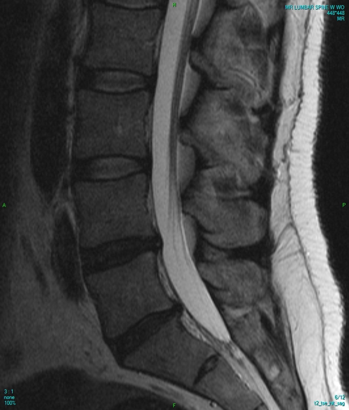
Can MRI Be Used to Detect Osteoporosis?
What MRI Shows
MRI (Magnetic Resonance Imaging) is a tool that can be used to visualise soft tissue and bone microarchitecture.
Unlike DEXA, which uses radiation, MRI relies on a powerful magnet and radio waves to scan the body. MRI scans are therefore safe and repeatable for most people6.
MRI can also assess much more than just bone density. It can be used to look at bone quality, which is assessed using Vertebral Bone Quality (VBQ) scores7.
What the Research Says
MRI shows promise for detecting bone changes earlier and more comprehensively than DEXA:
- Studies show MRI may detect bone deterioration before BMD drops, and can identify structural weakness even when DEXA results are normal. In a study of post-menopausal women not considered to have osteoporosis (i.e., BMD T-score over -2.5), those with fractures showed significant deterioration to bone tissue, such as 17 per cent higher erosion compared to those without fractures8. Therefore, MRI accurately identified microstructural issues that are suggestive of diminished bone health even when the DEXA scale indicated otherwise.
- MRI offers the potential to better predict fracture risk. Research shows that microstructural information derived from MRI outperforms BMD alone when assessing fracture risk9.
- Advancements in MRI technology offer more accurate bone imaging. Ultrashort Echo Time MRI (UTE-MRI) is an emerging technique that offers highly precise imaging of bone microstructure and could offer a non-invasive way to accurately detect osteoporosis in patients10,11.
Challenges and Future Potential
Cost and Accessibility
MRI is not yet the gold standard for osteoporosis detection. The cost and accessibility of MRI make the use of this technique challenging:
- Cost: MRI is more expensive than DEXA
- Accessibility: MRI is less widely available in general practice, with its use mostly localised to research and specialist centres
Not Yet Standard Practice
The challenges associated with MRI mean that there are no official UK guidelines recommending MRI as a diagnostic tool for osteoporosis. More longitudinal research and cost-benefit analysis are needed before this technique can rival DEXA.
Promising Outlook
As MRI technology becomes faster and more accessible, its role in the early detection of osteoporosis could expand. It could benefit those under 50 or with a strong family history, where DEXA scans are not suitable.
Just because MRI is not an officially recognised diagnostic tool for osteoporosis does not mean it is inaccessible. Ezra offers MRI screenings that are valuable not only to determine bone health but also multi-organ health. Proactive MRI scans can help detect osteoporosis and other conditions sooner.
Summary: A Step Towards Earlier, Smarter Detection
DEXA is the industry standard for the detection of osteoporosis. While MRI may not replace DEXA in the near future, it shows promise in identifying bone changes sooner. Bone density is not the only indicator of bone health, and MRI offers a way to analyse both bone density and bone quality.
Many individuals with osteoporosis go undiagnosed. Fractures don’t just affect your health; they also impact your level of independence and quality of life. Early detection is key to making lifestyle and health changes that can prevent fractures from ever occurring. For health-conscious adults, a multi-organ MRI scan may reveal more than just cancer risk, it could offer insight into bone health, too.Want to stay ahead of silent conditions like osteoporosis? The Ezra MRI scan with Spine covers up to 14 organs and provides early insights across multiple health areas. Book your scan today.
Understand your risk for cancer with our 5 minute quiz.
Our scan is designed to detect potential cancer early.
References
1. NHS. Osteoporosis. NHS inform. October 2025. Accessed December 12, 2025. https://www.nhsinform.scot/illnesses-and-conditions/muscle-bone-and-joints/conditions-that-can-affect-multiple-parts-of-the-body/osteoporosis/
2. NHS. Osteoporosis. NHS. October 3, 2022. Accessed December 9, 2025. https://nhsuk-cms-fde-prod-uks-dybwftgwcqgsdmfh.a03.azurefd.net/conditions/osteoporosis/
3. Baji P, Patel R, Judge A, et al. Organisational factors associated with hospital costs and patient mortality in the 365 days following hip fracture in England and Wales (REDUCE): a record-linkage cohort study. Lancet Healthy Longev. 2023;4(8):e386-e398. doi:10.1016/S2666-7568(23)00086-7
4. NHS. Osteoporosis - Treatment. NHS. April 4, 2018. Accessed December 12, 2025. https://nhsuk-cms-fde-prod-uks-dybwftgwcqgsdmfh.a03.azurefd.net/conditions/osteoporosis/treatment/
5. Williams S, Khan L, Licata AA. DXA and clinical challenges of fracture risk assessment in primary care. Cleve Clin J Med. 2021;88(11):615-622. doi:10.3949/ccjm.88a.20199
6. Ross J. MRI Scan: Uses, procedure, what to expect | Bupa UK. Bupa. January 31, 2023. Accessed December 12, 2025. https://www.bupa.co.uk/health-information/surgery-and-procedures/mri-scan
7. Najafi A, Bagheri AB, Hadavi D, Mobedi A, Azarsina S, Chaghamirzayi P. Vertebral bone quality score as a new tool for osteoporosis diagnosis in patients undergoing lumbosacral fusion surgery: a single center cohort study. Eur J Transl Myol. 2024;34(4). doi:10.4081/ejtm.2024.12311
8. Kijowski R, Tuite M, Kruger D, Munoz Del Rio A, Kleerekoper M, Binkley N. Evaluation of trabecular microarchitecture in nonosteoporotic postmenopausal women with and without fracture. J Bone Miner Res. 2012;27(7):1494-1500. doi:10.1002/jbmr.1595
9. Sollmann N, Löffler MT, Kronthaler S, et al. MRI-Based Quantitative Osteoporosis Imaging at the Spine and Femur. J Magn Reson Imaging. 2021;54(1):12-35. doi:10.1002/jmri.27260
10. Jerban S, Moazamian D, Mohammadi HS, et al. More accurate trabecular bone imaging using UTE MRI at the resonance frequency of fat. Bone. 2024;184:117096. doi:10.1016/j.bone.2024.117096
11. Sollmann N, Löffler MT, Kronthaler S, et al. MRI-Based Quantitative Osteoporosis Imaging at the Spine and Femur. J Magn Reson Imaging. 2021;54(1):12-35. doi:10.1002/jmri.27260


