Conditions our MRI scans have found
Fibroid degeneration
Uterine fibroids (also called uterine leiomyomas or myomas) are abnormal growths in the muscle of the uterus. Fibroids do not generally have malignant (cancerous) potential, but can occasionally be the cause of some types of abdominal pain or prolonged/irregular menses.Blood vessels supply uterine fibroids with the nutrients and oxygen they need to grow. Uterine fibroid degeneration occurs when a fibroid outgrows its limited blood supply. The connecting blood vessels can no longer provide enough oxygen and nutrients to the fibroid causing its cells to die, or degenerate.
Free fluid in the pelvis (biologically female)
Small amounts of free fluid in the pelvis is a normal finding in pre- and post-menopausal women without symptoms or known abdominal/pelvic disease. However, larger amounts of free fluid can be due to a pathological process including bleeding into the stomach, trauma, acute inflammation, rigorous exercise (e.g. running a marathon), and gastritis.
Fluid in the inguinal canal
The inguinal canal is a passage in the abdominal wall near the groin. It serves as a pathway through which structures (spermatic cords in males and the round ligament of the uterus in females) can pass from the abdominal wall to the external genitalia. There is one inguinal canal in each groin. The inguinal canals are larger and more prominent in males. Fluid within the inguinal canal may be associated with inguinal hernias (when tissue, such as part of the intestine or fat, pushes through a weak spot in the abdominal wall).
Fat - containing lesion of the femur
Fat-containing tumors are the most common soft-tissue tumors encountered clinically with the vast majority being benign (non-cancerous). Lipomas are the most common type of fat-containing tumors and demonstrate a characteristic appearance on MRI. The cause is not fully understood, but there is a genetic factor as they typically run in families. Usually these tumors do not cause symptoms, but can become painful if large and pressing on nearby nerves. Diagnostic evaluation includes physical exam, biopsy (tissue sampling), and a dedicated CT or MRI. Treatment is not indicated unless the tumor is growing in size or causes pain, in which case it can be removed surgically. Fat-containing tumors may also resemble a form of cancer called liposarcoma (cancerous tumors in fatty tissues that grow rapidly, don’t move under the skin if pushed from side to side, and are usually painful). A biopsy and/or MRI or CT scan is typically performed if liposarcoma is suspected.
Fibrocystic changes of the femur
There is a spot (lesion) on your femur (thigh bone) that looks like a collection of fibers and cysts (i.e. fibrocystic). When it is located in this part of the femur, it is also called an impingement cyst. Impingement cysts can be associated with a syndrome called femoroacetabular impingement (FAI) where the femur is too tight against the hip, causing increased friction with hip movements.
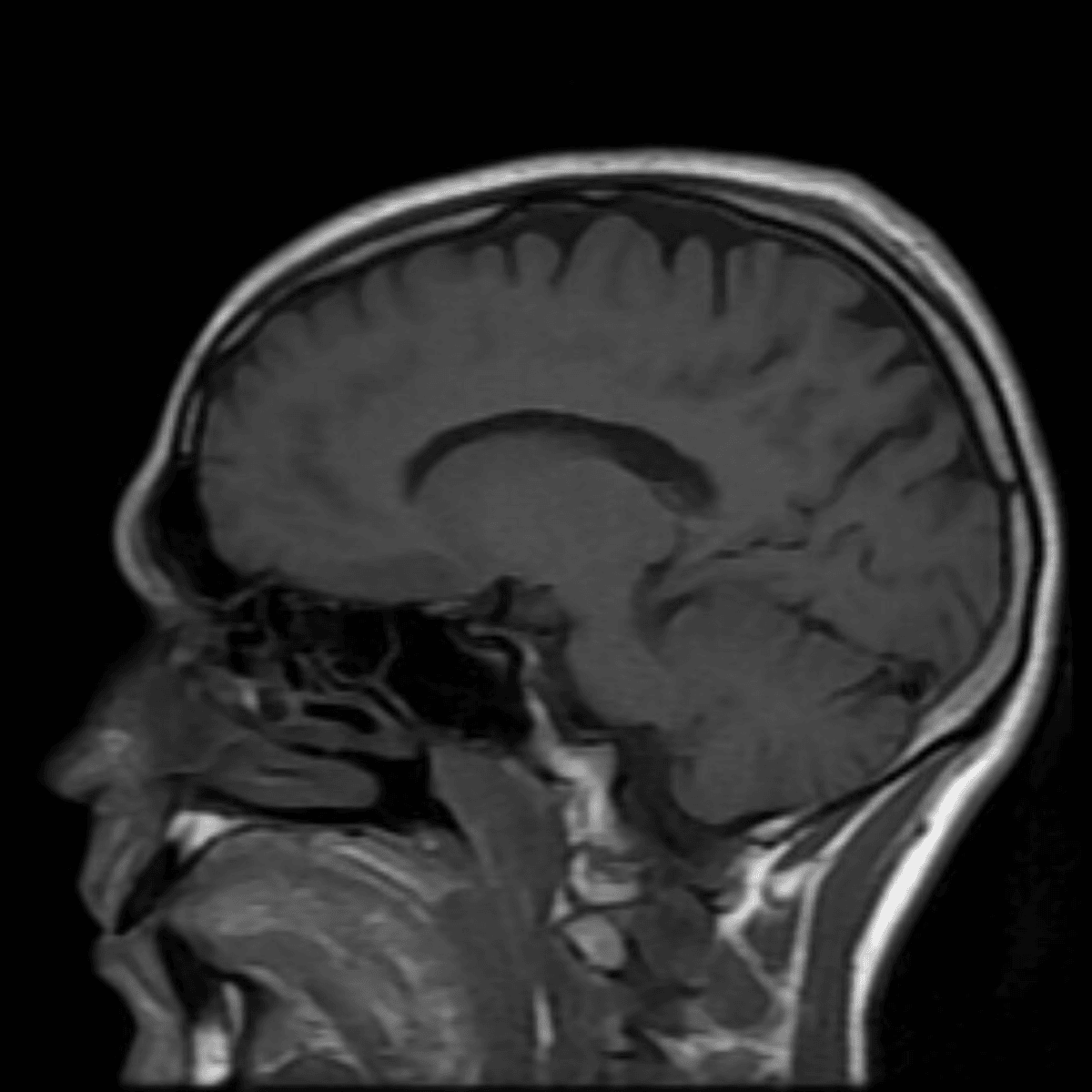
Focus of signal abnormality
A localized area (i.e. focus) that appears abnormal on MRI brain images. This finding is nonspecific, meaning it is difficult to say what caused it.
Can't find what you're looking for?
Frequently Asked Questions
Unfortunately at this time, we are unable to scan people with pacemakers. There is a risk that the MRI magnetic fields will disrupt its operation, and we don't want that.
There are many different types of implants. We will need to know more about what you have. The good news is that almost all implants are MRI-safe. There is a chance the implant will affect the images we can get from the surrounding tissues.
Yes. Almost all IUDs are MRI-safe. Regardless, we check the MRI safety of all devices. Common MRI-safe IUDs are the Mirena and the Copper T.
Yes you can. There will be extra precautions we will take to ensure your safety while in the machine, so please inform staff before entering the MRI.
Yes. This is completely safe.