MRI for Gallbladder Cancer Detection
Gallbladder cancer is a rare but aggressive cancer that starts in the gallbladder, a small pear-shaped organ located beneath the liver and responsible for storing bile for digestion. Globally, it accounts for about 1.2 per cent of all cancer diagnoses1.
Gallbladder MRI is a non-invasive imaging tool that uses magnetic fields and radio waves to produce detailed images of the gallbladder and surrounding ducts, aiding in diagnosing conditions like gallstones, inflammation, and tumours2. Magnetic Resonance Cholangiopancreatography (MRCP), a specialised type of gallbladder MRI, is particularly effective for investigating bile duct diseases and differentiating between benign and malignant conditions.
Why You Might Need a Gallbladder MRI
A gallbladder MRI might be needed to evaluate unexplained upper abdominal pain or jaundice, to detect cancer or masses, when other scans are inconclusive, and to differentiate benign from malignant gallbladder conditions.
Evaluating Unexplained Pain or Jaundice
- MRI is commonly ordered when patients have upper abdominal pain or jaundice, and preliminary tests (labs, ultrasound) do not clarify the cause.
- The scan can identify hidden gallstones, biliary tract infections, pancreatitis, bile duct obstructions, or other structural abnormalities.
Detecting Cancer, Polyps, or Masses
- Gallbladder MRI provides detailed images helpful for identifying tumours, polyps, and masses that might not be visible on other imaging tests3.
- MRI scans can distinguish the size, shape, and spread of lesions, which is essential for cancer detection and surgical planning4.
When Other Imaging is Inconclusive
- Ultrasound and CT are often the first-line investigations, but if results are unclear or the area cannot be visualised well (e.g., due to patient body habitus or overlying bowel gas), MRI is recommended for clearer, non-invasive imaging5.
- Specialised MRI (MRCP) is particularly useful for evaluating the bile ducts, where conventional ultrasound may not reach3.
Differentiating Benign from Malignant Conditions
- MRI can help clinicians distinguish benign conditions like adenomyomatosis or chronic cholecystitis from malignancies by analysing specific image features (layered patterns, signal intensity, papillary growth, enhancement patterns)4,6.
- Studies show that MRI features such as diffusion patterns and enhancement characteristics have high diagnostic accuracy; combinations of these features can strongly suggest malignancy.
How to Prepare for Your Gallbladder MRI
Here are a few tips to help you prepare for your MRI7:
- Take your usual medications and eat normally, unless instructed to fast for sedation or contrast. Avoid heavy caffeine intake and stay hydrated.
- Inform staff about any metal implants or devices, and bring safety cards for “MR-Conditional” implants, such as pacemakers or aneurysm clips.
- Remove all metal items, including jewellery, hairpins, dental plates, and transdermal patches with foil, to prevent image distortion.
- Wear comfortable, metal-free clothing; ask about earplugs, music, or mirror goggles if you’re claustrophobic.
- If contrast is planned, fast for 4–6 hours prior to the procedure. Inform staff about any kidney issues or past reactions to contrast agents, as safer alternatives may be available.
- Arrange an escort home if you need sedation for claustrophobia.
You can read more about preparation for Ezra’s Full Body Scan here.
What Happens During the Gallbladder Scan?
Upon arrival for your MRI, you will need to check in and complete a screening form. This will allow you to confirm the presence of implants, allergies, and whether you might need any anxiety medication.
During the scan, you will lie down on a sliding table. A dedicated surface or phased-array coil is typically placed over the limb or region of interest8. Your head will be nestled in a small cushion that will keep you still. The scan typically lasts 30-45 minutes of actual “table time”, during which the technician may acquire multiple sequences (settings). Expect loud knocking noises (up to 110 dB); earplugs or headphones are provided to reduce discomfort. It’s normal to feel mild table vibrations.
You’ll stay in touch with the team via a two-way intercom and a squeeze bulb, allowing you to communicate or pause the scan if needed. If contrast is required, it’s injected halfway through, possibly causing a brief cool sensation. After the final sequence, the coil is removed, and you’re free to go.
At Ezra, our Full Body Plus scan takes around 60 minutes total, with 45 minutes of table time. Earplugs or headphones are available.
After the scan, you will be contacted by a medical provider working with Ezra within roughly a week. On the day of the appointment, you will receive a copy of your report and access to your scanned images through the online portal.
MRI Safety, Risks, & Side Effects
MRI is generally considered very safe when proper screening and protocols are followed, but certain risks and side effects should be understood:
- Metal and implants: The strong 3-Tesla magnet can pull or heat older pacemakers, aneurysm clips, or metal fragments9. Most modern “MR-Conditional” devices (like cochlear implants or pain pumps) are safe after screening, but all implants must be checked before scanning10.
- Gadolinium contrast: Macrocyclic gadolinium agents (e.g., gadobutrol) have an extremely low risk of allergic reactions or nephrogenic systemic fibrosis (NSF) when kidneys are healthy11. However, gadolinium can accumulate in tissues, and rare side effects such as headaches or skin changes have been reported. Many centres now offer contrast-free alternatives for routine follow-up12. You can read more about gadolinium contrast side effects here.
- Incidental findings: Incidental gallbladder polyps are detected in approximately 3-6 per cent of the general population undergoing imaging13.
- Claustrophobia: Anxiety inside the scanner is common. Wide-bore scanners, music, mirror goggles, or a single dose of oral sedative can help alleviate symptoms. Open MRI is an option if image detail can be sacrificed.
- Zero Ionising Radiation: MRI uses magnetic fields and radio waves, not X-rays, so there is no ionising radiation exposure, making it safer for repeated scans compared to CT scans14.
- Minor Sensations: Expect loud knocking, mild table vibration, and a brief cool flush if contrast is injected. Rare side effects include headaches, fatigue, or mild skin heating.
A deeper dive into possible side effects (such as heat, headaches, and gadolinium deposition) is available in our full guide.
At Ezra, we employ a contrast-free approach using wide-bore T3 machines to deliver a comfortable scanning experience.
Terms You Might See in Your MRI Report (And What They Mean)
MRI reports of the gallbladder include specific terms that help clinicians assess the nature of a lesion or condition. Some common terms (and their meanings) include:
Focal Wall Thickening: This indicates a localised increase in gallbladder wall thickness, which may indicate a possible cancerous lesion15.
Rokitansky-Aschoff Sinuses (RAS): Represent tiny pockets within the gallbladder wall that are considered to be a benign finding16.
Diffusion Restriction: Describes tissues that impede water movement, often suggesting malignancy due to increased cellular density.
Enhancement Pattern: Refers to how a lesion lights up after contrast is given, revealing details about its behaviour and aggressiveness.
Mass Effect: Indicates that a lesion is pushing on or displacing nearby structures, showing its size or potential impact.
Ezra provides a radiologist-reviewed report in a non-technical and easy-to-understand format on your dashboard.
After the MRI Scan
After the MRI scan, you will be free to go home and continue with your day without any precautions17. If you received a sedative, you will need another person to pick you up. You will also not be able to drive, consume alcohol, or operate heavy machinery 24 hours after the sedative.
A team of experts will review your results and determine whether a follow-up is necessary and recommend the appropriate treatment if needed. If abnormalities are found, you may undergo ongoing monitoring every 2-3 months to track recurrence. You can receive support in the form of counselling and advice on how to handle aspects like claustrophobia.
If you have a scan with us here at Ezra, you will receive your report within five to seven days and have the option to discuss it with a medical practitioner. You can also access your scan images through the online portal.
What MRI Can Show About Gallbladder Cancer
MRI is a vital tool for diagnosing and staging gallbladder cancer, capable of revealing distinct patterns such as a mass replacing the gallbladder, thickened or irregular wall structures, liver invasion, lymph node enlargement, and bile duct obstruction, often outperforming ultrasound and CT for early-stage or complex cases18–20.
Mass Replacing the Gallbladder
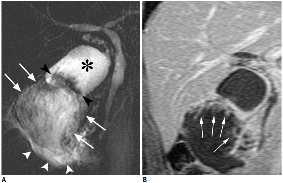
MRI can show a large, solid tumour that occupies or completely replaces the gallbladder; this is the most common imaging presentation and often indicates advanced disease. Such masses usually invade adjacent organs, particularly the liver, and may contain calcifications or gallstones within the tumour18,19,21,22.
Thickened or Irregular Wall Patterns
Malignant thickening of the gallbladder wall on MRI typically appears as diffuse, nodular thickening with disrupted mucosal lines and absence of the normal layered appearance; apparent diffusion coefficient (ADC) values can help differentiate malignant from benign causes, with malignancy showing low ADC values and early contrast enhancement15,23,24.
Invasion into Liver and Surrounding Tissues
MRI provides clear visualisation of local and advanced invasion, especially into the liver, using hepatobiliary contrast agents and dynamic scanning, often delivering higher sensitivity than other modalities; delayed enhancement in adjacent tissues can indicate hepatic involvement18,20,25.
Lymph Node Enlargement and Bile Duct Obstruction
MRI, particularly when combined with MRCP, is sensitive for detecting lymph node enlargement and obstructive jaundice caused by tumour involvement of lymphatics or the biliary tree, aiding staging and prognosis. Enlarged lymph nodes in regions like the hepatic hilum are common and can be evaluated by MRI, though smaller nodes may be missed2,19,26.
Early-Stage Lesions Missed by Ultrasounds or CT
MRI’s superior soft tissue characterisation allows for the detection of small, early-stage gallbladder lesions that might be overlooked by ultrasound or computed tomography (CT), including small intraluminal masses or subtle wall thickening. This makes MRI especially valuable for comprehensive assessment in complex or inconclusive cases2,15,18,20.
Types of Gallbladder Tumours and How They Look on MRI
Gallbladder tumours exhibit distinct MRI characteristics depending on their type, helping differentiate malignant from benign conditions and metastasis from primary tumours.
- Adenocarcinoma is the most common gallbladder cancer, usually forming a solid mass showing restricted diffusion and irregular or heterogeneous enhancement after contrast. These masses often replace the gallbladder, infiltrate surrounding tissue, and exhibit low-to-intermediate T1 and high T2 signal intensity, sometimes with patchy necrosis or engulfed gallstones18,19,22,27.
.png)
- Mucinous carcinoma is a rare variant producing large amounts of mucin, giving rise to heterogeneous high T2 signals within the lesion. MRI may reveal linear or curvilinear hypointense “mucus thread” features on MRCP, differentiating mucin from solid tumour components or stones28. Gadolinium-enhanced imaging accentuates papillary projections and mucus threads that do not enhance28.
- Metastasis (such as from melanoma or other cancers) creates highly vascular masses that often mimic primary tumours, display arterial enhancement, and may be hyperintense on T1-weighted images due to melanin content. These lesions generally attach to the wall and can cause ductal dilation if they obstruct the biliary tree29.
- Benign Mimics like adenomyomatosis are a common benign condition marked by focal or diffuse gallbladder wall thickening with intramural cystic spaces, seen as the “pearl necklace sign” on T2-weighted and MRCP images. These represent Rokitansky-Aschoff sinuses (RAS) and give high T2 signal foci within the wall, highly specific for adenomyomatosis, whereas serosal discontinuity suggests malignancy30,31.
Ezra screens for over 500 conditions, including the brain.
Types of MRI Scans Used in Gallbladder Cancer Detection
There are multiple types of MRI scans, all using different methods to give a better visualisation of the gallbladder.
- T1-weighted imaging provides detailed anatomical views of the gallbladder and its boundaries with the liver, helping to identify tumour presence and infiltration32.
- T2-weighted imaging excels at highlighting fluid-rich lesions and mucin within tumours, allowing for better detection of cystic or mucinous cancers33.
- Diffusion-weighted imaging (DWI) pinpoints tumour areas by revealing regions with restricted water movement due to high cellularity, which is typical of malignancy34.
- MRCP visualises the bile ducts non-invasively, showing ductal obstructions or tumour-related extensions into the biliary system35.
- Dynamic contrast-enhanced MRI evaluates how lesions absorb contrast over time, helping differentiate vascular tumour types from benign conditions based on their unique enhancement patterns22,33.
MRI vs. Other Imaging Tests for Gallbladder Cancer Detection
Cost
At Ezra, our MRI Scan (which includes the head, neck, abdomen, and pelvis) is offered at a £1495 all-inclusive price, including a 45-minute follow-up review of your scan findings with a medical practitioner. We have locations in London and Sidcup, with further locations across the UK coming soon.
Frequently Asked Questions
Can MRI detect early gallbladder cancer?
Yes, MRI can detect gallbladder cancer with high sensitivity, particularly when advanced imaging protocols are used.
Is the scan painful?
No, MRI scans are painless and non-invasive.
Do I need contrast for a gallbladder MRI?
Contrast is often recommended for a gallbladder MRI to achieve the most accurate assessment, but some diagnostic information can be obtained without it.
Can MRI replace a biopsy?
No, MRI cannot replace a biopsy, as tissue diagnosis is still required to confirm gallbladder cancer.
Key Takeaways
- Gallbladder MRI is essential for accurate cancer detection, staging, and guiding treatment decisions.
- MRI provides superior soft tissue detail compared to ultrasound or CT, allowing for clearer visualisation of tumours and surrounding anatomy.
- Specific MRI patterns help differentiate benign from malignant gallbladder lesions, improving diagnostic confidence and patient outcomes.
- Proper patient preparation and understanding the terminology in MRI reports can enhance the care journey and empower informed medical decisions.


References
1. Rawla P, Sunkara T, Thandra KC, Barsouk A. Epidemiology of gallbladder cancer. Clin Exp Hepatol. 2019;5(2):93-102. doi:10.5114/ceh.2019.85166
2. Tests for gallbladder cancer. Accessed August 25, 2025. https://www.cancerresearchuk.org/about-cancer/gallbladder-cancer/getting-diagnosed/tests-gallbladder-cancer
3. Radiology (ACR) RS of NA (RSNA) and AC of. Gallstones. Radiologyinfo.org. Accessed August 25, 2025. https://www.radiologyinfo.org/en/info/gallstones
4. He WW, Zhu JG, Pylypenko D, et al. Differentiating benign from malignant gallbladder wall thickening in non-contrast MRI imaging: Preliminary study of a combined diagnostic indicator. Medicine (Baltimore). 2022;101(40):e30861. doi:10.1097/MD.0000000000030861
5. Mendelson R. Imaging for chronic abdominal pain in adults. Aust Prescr. 2015;38(2):49-54. doi:10.18773/austprescr.2015.019
6. Yoshimitsu K, Honda H, Kaneko K, et al. Dynamic MRI of the gallbladder lesions: differentiation of benign from malignant. J Magn Reson Imaging. 1997;7(4):696-701. doi:10.1002/jmri.1880070415
7. Radiology (ACR) RS of NA (RSNA) and AC of. Magnetic Resonance Imaging (MRI) - Head. Radiologyinfo.org. Accessed July 3, 2025. https://www.radiologyinfo.org/en/info/mri-brain
8. Gruber B, Froeling M, Leiner T, Klomp DWJ. RF coils: A practical guide for nonphysicists. J Magn Reson Imaging. 2018;48(3):590-604. doi:10.1002/jmri.26187
9. Gill A, Shellock FG. Assessment of MRI issues at 3-Tesla for metallic surgical implants: findings applied to 61 additional skin closure staples and vessel ligation clips. J Cardiovasc Magn Reson. 2012;14(1):3. doi:10.1186/1532-429X-14-3
10. Potential Hazards and Risks. UCSF Radiology. January 20, 2016. Accessed March 14, 2025. https://radiology.ucsf.edu/patient-care/patient-safety/mri/potential-hazards-risks
11. Costello JR, Kalb B, Martin DR. Incidence and Risk Factors for Gadolinium-Based Contrast Agent Immediate Reactions. Top Magn Reson Imaging. 2016;25(6):257-263. doi:10.1097/RMR.0000000000000109
12. McDonald RJ, McDonald JS, Kallmes DF, et al. Gadolinium Deposition in Human Brain Tissues after Contrast-enhanced MR Imaging in Adult Patients without Intracranial Abnormalities. Radiology. 2017;285(2):546-554. doi:10.1148/radiol.2017161595
13. Kamaya, A., Fung, C., Szpakowski, J-L., et al. Management of Incidentally Detected Gallbladder Polyps: Society of Radiologists in Ultrasound Consensus Conference Recommendations. Radiology. 2022;000:1-12. doi: 10.1148/radiol.213079
14. Mall MA, Stahl M, Graeber SY, Sommerburg O, Kauczor HU, Wielpütz MO. Early detection and sensitive monitoring of CF lung disease: Prospects of improved and safer imaging. Pediatr Pulmonol. 2016;51(S44):S49-S60. doi:10.1002/ppul.23537
15. Gupta P, Marodia Y, Bansal A, et al. Imaging-based algorithmic approach to gallbladder wall thickening. World Journal of Gastroenterology. 2020;26(40):6163. doi:10.3748/wjg.v26.i40.6163
16. Golse N, Lewin M, Rode A, Sebagh M, Mabrut JY. Gallbladder adenomyomatosis: Diagnosis and management. Journal of Visceral Surgery. 2017;154(5):345-353. doi:10.1016/j.jviscsurg.2017.06.004
17. MRI scan. NHS inform. Accessed July 3, 2025. https://www.nhsinform.scot/tests-and-treatments/scans-and-x-rays/mri-scan/
18. Neculoiu D, Neculoiu LC, Popa RM, Manea RM. The Many Hidden Faces of Gallbladder Carcinoma on CT and MRI Imaging—From A to Z. Diagnostics (Basel). 2024;14(5):475. doi:10.3390/diagnostics14050475
19. Radswiki T. Gallbladder carcinoma | Radiology Reference Article | Radiopaedia.org. Radiopaedia. doi:10.53347/rID-12407
20. Hwang J, Kim YK, Choi D, et al. Gadoxetic acid-enhanced MRI for T-staging of gallbladder carcinoma: emphasis on liver invasion. Br J Radiol. 2014;87(1033):20130608. doi:10.1259/bjr.20130608
21. Ramachandran A, Srivastava DN, Madhusudhan KS. Gallbladder cancer revisited: the evolving role of a radiologist. Br J Radiol. 2021;94(1117):20200726. doi:10.1259/bjr.20200726
22. Furlan A, Ferris JV, Hosseinzadeh K, Borhani AA. Gallbladder Carcinoma Update: Multimodality Imaging Evaluation, Staging, and Treatment Options. American Journal of Roentgenology. 2008;191(5):1440-1447. doi:10.2214/AJR.07.3599
23. Lopes Vendrami C, Magnetta MJ, Mittal PK, Moreno CC, Miller FH. Gallbladder Carcinoma and Its Differential Diagnosis at MRI: What Radiologists Should Know. Radiographics. 2021;41(1):78-95. doi:10.1148/rg.2021200087
24. van Breda Vriesman AC, Engelbrecht MR, Smithuis RHM, Puylaert JBCM. Diffuse Gallbladder Wall Thickening: Differential Diagnosis. American Journal of Roentgenology. 2007;188(2):495-501. doi:10.2214/AJR.05.1712
25. Choi J, Kim HJ, Jang SK, Paik SY, Kim KH. Synchronous Cancers of Hepatic Angiosarcoma and Gallbladder Adenocarcinoma, Mimicking Gallbladder Cancer with Hepatic Invasion: a Case Report. Investigative Magnetic Resonance Imaging. 2020;24(2):90-94. doi:10.13104/imri.2020.24.2.90
26. Li Y, Song Y, Zhang Y, Liu S. Progress in gallbladder cancer with lymph node metastasis. Front Oncol. 2022;12:966835. doi:10.3389/fonc.2022.966835
27. Wagreich JM, Shapiro RS, Glajchen N, Seijo L. Mri findings in adenosquamous carcinoma of the gallbladder. Clinical Imaging. 1998;22(2):130-133. doi:10.1016/S0899-7071(97)00079-X
28. Huang CP, Chiou YY, Chou YH, Chiang JH, Chang CY. Imaging Findings in Mucin-producing Carcinoma of the Gallbladder. Journal of the Formosan Medical Association. 2006;105(5):427-430. doi:10.1016/S0929-6646(09)60141-8
29. Cocco G, Delli Pizzi A, Basilico R, et al. Imaging of gallbladder metastasis. Insights Imaging. 2021;12:100. doi:10.1186/s13244-021-01049-8
30. Haradome H, Ichikawa T, Sou H, et al. The pearl necklace sign: an imaging sign of adenomyomatosis of the gallbladder at MR cholangiopancreatography. Radiology. 2003;227(1):80-88. doi:10.1148/radiol.2271011378
31. Riddell ZC, Corallo C, Albazaz R, Foley KG. Gallbladder polyps and adenomyomatosis. Br J Radiol. 2023;96(1142):20220115. doi:10.1259/bjr.20220115
32. Mitchell CH, Johnson PT, Fishman EK, Hruban RH, Raman SP. Features Suggestive of Gallbladder Malignancy. J Comput Assist Tomogr. 2014;38(2):235-241. doi:10.1097/RCT.0b013e3182aafb6b
33. Han S, Lee YH, Kim YR, Soh EG. Usefulness of MRI Scoring System for Differential Diagnosis between Xanthogranulomatous Cholecystitis and Wall-Thickening Type Gallbladder Cancer. Journal of the Korean Society of Radiology. 2024;85(1):147-160. doi:10.3348/jksr.2023.0036
34. Ohki K, Igarashi T, Yakabe H, et al. Differentiating gallbladder cancer from polyps using non-enhanced magnetic resonance imaging. Pol J Radiol. 2024;89:e106-e114. doi:10.5114/pjr.2024.135730
35. Al-Dhuhli H. Role of Magnetic Resonance Cholangiopancreatography in the Evaluation of Biliary Disease. Sultan Qaboos Univ Med J. 2009;9(3):341-352.
36. Wood J. Private MRI scan costs explained. Practice Plus Group. December 13, 2023. Accessed January 15, 2025. https://practiceplusgroup.com/knowledge-hub/private-mri-scan-costs-explained/